My first experiences with AT ELANA 841P and ARTEVO 850

This article by Liem Trinh, MD highlights his experiences with the AT ELANA® 841P hydrophobic trifocal IOL and the ARTEVO® 750 and ARTEVO® 850 microscopes from ZEISS. Dr. Trinh discusses the benefits of the trifocal design of the AT ELANA 841P IOL and its ability to provide spectacle independence at all distances after cataract surgery. He also shares his positive experiences with the ARTEVO 850 microscope, which offers improved surgical visualization with RGB LED illumination and 3D viewing on a high dynamic range monitor. Additionally, Dr. Trinh discusses the potential benefits of integrated intraoperative optical coherence tomography (OCT) and the redesigned CALLISTO eye user interface.
Founded in the 13th century, the Quinze-Vingts National Ophthalmology Hospital in Paris is commonly noted to be one of the oldest hospitals in France, but it is known to be equipped with the best in modern diagnostic and surgical technology. I truly appreciate having access to the latest advances in equipment and software that elevate my performance as a surgeon and serve to enhance outcomes for my patients. And, as a self-described geek I am always eager to trial new technology. Therefore, the launch of the AT ELANA® 841P hydrophobic trifocal IOL and the introduction of the ARTEVO® 750 and ARTEVO® 850 microscopes from ZEISS were exciting developments for me. After learning about the features of these new products, I looked forward to using them, and in my first real-world clinical experiences they are fulfilling my expectations.
ZEISS AT ELANA 841P
Optical designs for presbyopia-correcting IOLs continue to evolve, but in my experience and according to findings of a recent meta-analysis, a trifocal optical design gives patients the best chance of achieving spectacle independence at all distances after cataract surgery.1 Going back to 2008, the first presbyopia-correcting IOL I implanted was the AT LISA® 809M (Carl Zeiss Meditec AG, Jena, Germany), a hydrophilic bifocal lens that provided high quality vision for distance and near, but often left patients needing correction for intermediate vision tasks. Thus, I was very happy when the trifocal AT LISA® tri 839MP became available because it combined the proven performance of the AT LISA platform with the functional benefit of a trifocal optic.
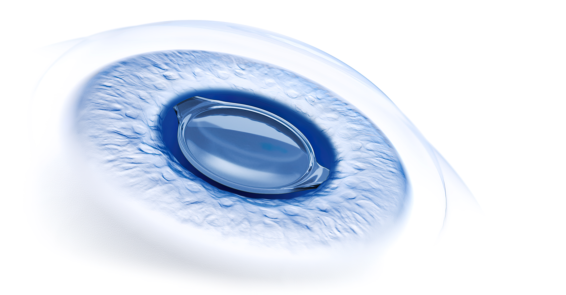
Now, with the AT ELANA 841P, the newest member of ZEISS IOLs, I can give my patients seeking presbyopia correction the advantages of a fully preloaded trifocal IOL with a glistening-free*hydrophobic acrylic biomaterial and heparin-coated** surface that ensures good optical clarity as well as smooth and controlled unfolding. Furthermore, the C-loop platform and step-vaulted, rigid optic-haptic junction found on the AT ELANA 841P facilitate centration and capsular contact to minimize posterior capsule opacification (PCO). The design of the AT ELANA 841P optic is based on the AT LISA tri 839MP, but it is modified to have increased light transmission efficiency and a higher allocation of light towards near vision to enhance near-to-intermediate vision without compromising far vision.***
Personal preferences
Different surgeons favor different IOL biomaterials, but compared to hydrophilic acrylic, hydrophobic acrylic material has the advantage of being associated with a lower incidence of PCO.2 I inject the AT ELANA 841P through a 2.4 mm incision. As a fully preloaded lens, the AT ELANA 841P contributes to surgical efficiency, and the lens unfolds gently and easily, enabling precise positioning.
The refractive and functional outcomes from my first use of the AT ELANA 841P have been as favorable as my initial intraoperative experience. While there is always uncertainty about achieving the targeted refractive outcome when implanting a new IOL with use of the manufacturer’s recommended IOL constant, my analysis of data from the first 10 patients who underwent bilateral implantation showed excellent accuracy. At 3 months after surgery, spherical equivalent averaged +0.09 D and was <1.0 D in all eyes; mean residual astigmatism was 0.86 D. Mean (decimal) monocular best corrected distance visual acuity was 0.9, all eyes had a best corrected intermediate visual acuity >0.7, and best corrected near visual acuity was >0.8 in 90% of eyes. The defocus curve for my patients was consistent with the published findings from bench tests that showed the AT ELANA841P has good optical performance across an extended range of distances.3
ARTEVO 750 and ARTEVO 850
I have been very happy operating with the OPMI LUMERA® 700 optical microscope because I find it provides high clarity images with excellent contrast and color. In addition, I appreciate the microscope’s connectivity with other ZEISS diagnostic, planning, and surgical platforms, which I find increases workflow efficiency.
Yet, when it comes to technology, it seems that there are infinite possibilities for innovation and advancements, and ZEISS proves that point again with the introduction of two new surgical microscopes – the ARTEVO 750, an optical microscope with traditional oculars, and the ARTEVO 850, a hybrid device with both a 3D monitor for digital visualization and traditional oculars.
I recently had the opportunity to trial the new ARTEVO 850. The image quality of the LUMERA 700 was already outstanding according to my experience, making the improvement I found with the new ARTEVO 850 microscope even more impressive. I personally found that the ARTEVO 850 improves surgical visualization with the new RGB LED illumination. Equipped with a mix of red, green, and blue LEDs, the RGB illumination technology allows surgeons to adjust light color temperature from warm to cold (3000K to 6000K), offering the opportunity to finetune the surgical view according to personal preferences. I also found this feature may help to highlight specific anatomical details. For example, I noticed that switching to a warmer temperature during capsulorhexis made the red reflex even more stunning, aiding visualization of the capsule edge.
The ARTEVO 850 captures the surgical image with 2 4K 3-Chip cameras and displays it in 3D with true colors on the system’s 55-inch high dynamic range (HDR) monitor. Viewing the surgical image on the 3D HDR monitor was an amazing experience for me because I actually felt immersed inside the eye. In my experience and as demonstrated by multiple studies, operating with a heads-up configuration while viewing the 3D monitor is more ergonomic and comfortable than bending over the microscope, and it also gives other OR personnel a real-time view of the surgical procedure. However, the ARTEVO 850 is also equipped with a Hybrid Mode that allows surgeons to view the surgical field through the microscope’s oculars at any time during the procedure with just a simple switch of a button.
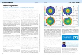
In addition, integrated intraoperative optical coherence tomography (OCT) is available as an optional add-on. In my opinion, the OCT image has excellent quality in high definition, and the OCT scan quality can be adjusted as desired. For example, after reducing the noise during a Descemet membrane endothelial keratoplasty case, I was able to better visualize the side of the graft (Figure 2). As another new feature, size indicators are displayed in the OCT image, and I am looking forward to applying this tool in Implantable Collamer Lens (ICL) cases so that I can estimate the size of the vault intraoperatively.
The ARTEVO 850 is digitally connected to the ZEISS Premium Cataract Workflow. Options for data overlays during cataract surgery include cataract assistance functions, e.g., for toric IOL alignment, and phaco parameters from QUATERA® 700. Along with the new ARTEVO 850 microscope, ZEISS introduced a redesigned CALLISTO eye user interface that I found to have a spectacular, intuitive touchscreen. Enhancing workflow efficiency, preoperative biometric data collected with the IOLMaster® 700 and planning data created using EQ Workplace® can be seamlessly transferred to the QUATERA 700 and the CALLISTO eye of the ARTEVO 850.
Conclusion
Innovation brings novelty to technology, but it must also provide value. Based on my early experience, the AT ELANA 841P IOL and ARTEVO 750 and ARTEVO 850 microscopes are true innovations offering important benefits that serve both me as a surgeon and my patients.
References
1. Li J, Sun B, Zhang Y, et al. Comparative efficacy and safety of all kinds of intraocular lenses in presbyopia-correcting cataract surgery: a systematic review and meta-analysis. BMC Ophthalmol. 2024;24(1):172.
2. Donachie PHJ, Barnes BL, Olaitan M, Sparrow JM, Buchan JC. The Royal College of Ophthalmologists' National Ophthalmology Database study of cataract surgery: Report 9, Risk factors for posterior capsule opacification. Eye (Lond). 2023;37(8):1633-1639.
3. Labuz G, Yan W, Khoramnia R, Auffarth GU. Optical-quality analysis and defocus-curve simulations of a novel hydrophobic trifocal intraocular lens. Clin Ophthalmol. 2023;17:3915-3923.
* Grade 1 (traces) or better for 85% of the patients up to and including 12 months according to Christiansen scale and based on internal clinical trial outcomes and on published clinical data.
** Fragment of heparin used in IOL surface coating with no pharmacological, immunological or metabolic action.
*** Compared to AT LISA tri in photopic conditions in virtual implantations and optical bench tests.
Liem Trinh, MD is an ophthalmologist at 15-20 National Ophthalmologic Hospital in Paris, France, specializing in refractive and cataract surgery. He is a paid consultant for Carl Zeiss Meditec.
Newsletter
Get the essential updates shaping the future of pharma manufacturing and compliance—subscribe today to Pharmaceutical Technology and never miss a breakthrough.


