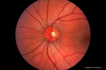
Clinical trials in wet AMD: Impact of central subfield thickness, volatility on visual acuity
Dr Justis P. Ehlers dissects the revelations from the data in the Phase 3 Hawk clinical trial regarding the impact of central subfield thickness, volatility and the overall impact on visual acuity.
In Fort Lauderdale, Florida, United States, Dr Justis P. Ehlers presented a talk entitled, “The impact of compartmental exudative volatility on visual acuity and ellipsoid zone integrity in neovascular AMD.”
Video transcript
So it's my pleasure to share some of the results from our research today. As many of you know, there's been a lot of work done recently examining the impact of central subfield thickness, volatility and the overall impact on visual acuity in clinical trials in wet AMD.
One of the big questions that remains is: What's driving that volatility in retinal thickness changes, and do those changes actually end up with important anatomical sequela that we can understand and strive to minimize as clinicians?
For this study, what we did is we looked at the Phase 3 Hawk clinical trial, we're able to use machine learning augmented, multilayer segmentation, as well as feature extraction to pull out specific fluid compartments including intraretinal fluid and subretinal fluid, as well as subretinal material.
Using this, we're able to look at volatility from week 12, after the loading phase all the way to the end of year one and to evaluate the impact of high volatility in each of those compartments on visual acuity outcomes and on anatomic sequela.
What we found was that each of those compartments intraretinal fluid, subretinal fluid, as well as subretinal material all had a negative impact on visual acuity if it was in the high volatility group, whereas eyes that were in the low volatility group continued to improve vision over time. When we looked at the anatomic sequela, what we found was, in fact, the subretinal fluid compartment had the biggest driver towards negative anatomic outcomes, particularly increase in subretinal fibrosis and subretinal material, as well as poor maintenance of ellipsoid zone integrity.
Interestingly, this flies a little bit in the face of what we know in terms of subretinal fluid. We've seen in the past that this can be linked with overall better visual acuity at specific time points, but what this shows is that potentially overall activity and the volatility of that compartment is an important discriminator between eyes that may do well with tolerating subretinal fluid, that static, versus really minimizing that subretinal fluid that may be volatile and dynamic. So overall, more work needs to be done in this area. It's really an exciting opportunity given the new and advanced image analysis techniques that we have, and we're excited to share it here at Retina World Congress.
Note: This transcript has been lightly edited for clarity.
Newsletter
Get the essential updates shaping the future of pharma manufacturing and compliance—subscribe today to Pharmaceutical Technology and never miss a breakthrough.


