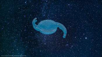
Toric intraocular lenses
Dr Monaco and colleagues performed a comparative study on two different toric lenses using wavefront technology for analysis. Here, he presents the findings.
Toric IOLs have been widely reported as effective, safe, and predictable,1–3 even in selected subgroups with high astigmatism.4 Yet, toric IOLs made of different materials, asphericity, toricity distribution, morphology and calculation algorithms could potentially have different outcomes.
Bearing this in mind, we planned to use wavefront technology in a comparative study to analyse the accuracy of preoperative calculation, precise intraoperative placement, and visual optical quality of two different toric IOLs.
By July 2011, 72 eyes had been enrolled to compare the results after implantation of Acrysof IQ SN6AT IOL (Alcon Laboratories Inc., Fort Worth, Texas, USA) and AT Torbi 709M (Carl Zeiss Meditec AG, Jena, Germany). The study included astigmatic patients having cataract surgery with IOL implantation in the capsular bag. Inclusion criteria were cataract; regular astigmatism; and no other ocular comorbidity that might influence visual outcome. Exclusion criteria were significant level of corneal higher-order aberrations (HOAs) (>0.350 µm) using the Zernike polynomials from the topography map, and a high irregular astigmatism index (>0.54, OPD-Scan II, Nidek Co. Ltd, Aichi, Japan).
Patients were randomly assigned to Group A (Acrysof) or Group B (AT Torbi) and implanted after a sutureless 2.2 mm coaxial phacoemulsification. Three months after surgery, evaluation of postoperative outcomes included analysis of visual acuity, wavefront refraction, spherical equivalent (SE), IOL axis, and optical quality. The objective optical quality of all surgical eyes was evaluated by analyzing (4.0 mm pupil diameter) the root mean square (RMS) of corneal, intraocular, and total HOAs Z(n,i) (3 ≤ n ≤ 8); coma Z(3,±1); trefoil Z(3,±2); and spherical aberration Z(4,0). The point-spread function (PSF) and modulation transfer function (MTF) were also obtained from total aberrations.
Postoperatively, no significant between-group differences were found in visual acuity and misalignment. The residual SE in Group A was significantly closer to emmetropia than that in Group B, which showed a mild myopic shift. The objective net refractive astigmatism magnitude was within ± 0.50 D of expected value at polar KP90 (polar value along 90-degree meridian) in 34 eyes (94.4%) in both groups. No statistically significant between-group difference was found in intraocular and total RMS of HOA, coma, trefoil, PSF, or the MTF. Intraocular and total spherical aberration Z(4,0) was statistically lower in Group B.
Conclusions
Despite the different morphology and calculation, in our comparative study,5 Acrysof and AT Torbi proved very similar as to clinical effectiveness in astigmatism correction and rotational stability. Eyes with Acrysof appeared significantly nearer to emmetropia, while AT Torbi induced significantly less spherical aberration.
Nonetheless, wavefront analysis added several interesting findings.
Even if spherical aberration with AT Torbi appeared to be statistically lower than that with Acrysof in intraocular and total aberrations, the different values did not appear to affect the markers of optical quality. There was no significant difference in the PSF and the MTF between groups and the distortion level of a simulated point source of light was very low for both IOLs.
The amount of HOAs generated by the IOLs was consistent with results in other studies despite using different aberrometers and pupil diameters.6–9
It is also possible to establish the toric IOL axis using the internal astigmatic wavefront map, where variation in dioptric power along a diameter chosen by the observer is plotted on a graph, as previously reported.10
References
1. A. Langenbucher et al., J. Refract. Surg., 2009;25:611–622.
2. J.D. Horn, Curr. Opin. Ophthalmol., 2007;18:58–61.
3. C. Novis, Curr. Opin. Ophthalmol., 2000;11:47–50.
4. N. Visser et al., J. Cataract Refract. Surg., 2011;37:1403–1410.
5. A. Scialdone, F. De Gaetano and G. Monaco, J. Cataract Refract. Surg., 2013;39:906–914.
6. H.P. Sandoval et al., Eye, 2008;22:1469–1475.
7. J.S. Pepose et al., Graefes Arch. Clin. Exp. Ophthalmol., 2009;247:965–973
8. S.W. Kim et al., Eye, 2008;22:1493–1498.
9. K. Hayashi, M. Yoshida and H. Hayashi, Eye, 2008;22:1476–1482.
10. A. Scialdone, G. Raimondi and G. Monaco, Eur. J. Ophthalmol., 2012;22:531–540.
Newsletter
Get the essential updates shaping the future of pharma manufacturing and compliance—subscribe today to Pharmaceutical Technology and never miss a breakthrough.


