
|Articles|November 1, 2017
Testing at point of care for improved patient satisfaction, re-treatment rates
After spending a few months testing for dry eye and treating the tear film before doing preoperative measurements, the re-treatment rate dropped from 11% to 3%.
Advertisement
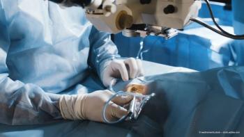
At our predominantly refractive surgery practice, my colleagues and I have always seen good refractive outcomes, but we found that a number of patients were unhappy. Surgery went perfectly and according to plan, and visual acuities were good, but patients were uncomfortable and frustrated with dry eye symptoms, such as grittiness, fluctuating vision, redness and foreign body sensation.
About two years ago, we began to look for ways to determine, through point-of-care preoperative testing, which patients were likely to experience this problem after surgery. Testing enables us to identify both symptomatic and asymptomatic ocular surface disease.
Asymptomatic patients who feel fine naturally do not think that they have a disease, but point-of-care testing offers them the proof they need to welcome treatment.
Once testing helps to pinpoint ocular surface disease before surgery, we can treat the problem and prevent patients from becoming unhappy with postoperative symptoms.
Importantly, dry eye treatment also allows us to obtain accurate preoperative measurements. If patients do not have a stable, good quality tear film, readings can be off by as much as 1.0 to 1.5 D. IOL calculations can be off as well.
We explain to patients with dry eye that we can perform the surgery they desire, but first we have to treat the ocular surface so we can plan surgery based on complete, accurate data.
A valuable series of tests
Dry eye testing begins subjectively. When patients check in at the practice, they fill out a questionnaire about their ocular symptoms. In response to the question, “What is your most bothersome symptom?” many patients offer answers that indicate dry eye.
Even patients who do not have symptoms can acquire dry eye after any surgery. However, if we do not detect, explain and treat the problem, the surgeon will be blamed.
Thus, my staff performs point-of-care testing for all patients. Test results determine whether the patient will have measurements for surgery that day or begin dry eye treatment and be measured later.
The first point-of-care clinical test we perform is a measurement of tear osmolarity (TearLab Osmolarity System, TearLab). It is important to do this test first because the effects of other tests on the ocular surface can influence the measurement.
Next, we test for the inflammatory marker MMP-9 (InflammaDry, Quidel) to determine if the disease has an inflammatory component. Thirdly, we measure changes in visual quality caused by an unstable tear film (HD Analyzer, Visiometrics).
The questionnaire and primary testing give a sense of what is happening on the ocular surface. For example, high scores on the dry eye questionnaire (Figure 1) and tear osmolarity above 308 mOsm/L or with an inter-eye difference above 8 mOsm/L indicate that the patient may have dry eye disease.
If meibomian gland dysfunction (MGD) is suspected, we can extend the workup to include meibography (LipiScan, TearScience). A noninvasive tear breakup time test with my three-dimensional Scheimpflug camera (Sirius, CSO) is also illuminating.
If the schedule does not build in time for meibography or other additional testing, we can reschedule a patient for further testing at a later time.
When the tests reveal dry eye, we are meticulous about improving the patient’s ocular surface before surgery. I prescribe anti-inflammatory eye drops, recommend omega-3 supplements and sometimes use punctal plugs to lower the outflow of tears from the eye.
When meibography shows significant atrophy or dilation of the glands, I start treatment with thermal pulsation gland expression (LipiFlow, TearScience) and/or intense pulsed light (IPL) treatment for inflammation (E-eye, E-Swin), as well as standard dry eye treatments, before measurements or surgery.
We perform dry eye testing at follow-up visits after surgery; patients continue dry eye treatment for six months.
Implementation and outcomes
The introduction of point-of-care dry eye testing required training and initially added time to the patient’s visit. Staff needed to get used to thinking about the importance of dry eye to our surgical outcomes and patients’ quality of life.
Training included how to perform the new tests, as well as establishment of tests technicians can perform before patients see the optometrist or the surgeon and those they should perform only when requested by a doctor. Once the tests became integrated into the practice workflow, they no longer added significant time to the patient’s visit.
The results were impressive. After we spent a few months testing for dry eye and treating the tear film before doing preoperative measurements, our re-treatment rate dropped from 11% to 3%. Our patient satisfaction questionnaires, which are filled out one month after surgery, showed significantly better scores.
More patients said they would recommend the procedure to family and friends. As a result, today, I would not think of performing any surgery without first testing for the presence of dry eye disease and treating the ocular surface.
Dr Erik L. Mertens, MD, FEBOphth
Dr Mertens is founder, medical director and eye surgeon at the Medipolis Multidisciplinary Clinic in Antwerp, Belgium. Dr Mertens is a consultant for Staar Surgical; Tearlab; Bausch&Lomb; Zeiss; PhysIOL; Ophtec; Rayner; Medicem; MST; and CapsuLaser.
Advertisement
Related Content
Advertisement


Latest CME


Advertisement
Advertisement