Retropupillary implantation of iris-claw lenses
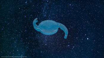
There are several surgical options available for the correction of aphakia, yet there is no clear winner in the competition for the best method: anterior chamber intraocular lenses (IOLs), suturing a posterior chamber IOL in the sulcus or using an iris claw lens. If an iris-claw lens, like the Verisyse IOL (AMO), is chosen, it will usually be fixated to the iris in the anterior chamber. Alternatively this IOL can also be fixated on the posterior surface of the iris (Figure 1).
In 1998, I carried out the first retropupillary implantation of an iris-claw lens in an aphakic and vitrectomized eye. These eyes always present a challenge because of their highly mobile iris diaphragm, which tries to move away when attempting to grasp the tissue for enclavation, particularly when performing regular anterior enclavation. To avoid this, I implanted the lens from the reverse side.
This technique, which I first reported in 2002,1 offers several advantages: it combines the benefits of posterior chamber implants with a low-risk method of surgery and it offers considerable cosmetic benefit, by hiding the IOL haptic and parts of the lens behind the iris. Retropupillary fixation of iris-claw lenses enhances stability, prevents tilting of the lens and reduces the glare phenomenon, which is characteristic of the lens being implanted in the anterior chamber. This method has been investigated further by several colleagues2,3,4,5 and is now frequently used.
How it's done, step-by-step
For the calculation of IOL power in this different position, an A-constant of 116.8 was estimated and used in the SRK II formula. Preoperatively, we tested pupil dilation and contractility using pilocarpine and cyclopentolate/ neosynephrine. The initial pupil size should be at least 5 mm, especially for surgeons who are not as familiar with the technique. Patients with larger iris defects, unregulated glaucoma, rubeosis iridis or iris atrophy should be excluded or handled with extreme care.
The lens is inserted vertically via a 5 mm scleral incision into the anterior chamber and then turned into a horizontal position. After sliding the IOL through the pupil to the retropupillary position, the lens is stabilized using specially designed forceps (H.P. Bräm AG, Switzerland) followed by an injection of Miochol (Novartis Ophthalmics) into the eye for miosis of the pupil.
When uniform contraction of the pupil is achieved the lens is lifted slightly so that the haptics can be recognized through the iris stroma visually as well as haptically. Via the paracenthesis, a spatula is used to press iris texture into the claws. The enclavation of the iris into the haptics is simple because of the natural pressure of the aqueous humour and because a peripheral iridotomy is not necessary. After aspiration of the ophthalmic viscosurgical device (OVD) (Healon; AMO) the scleral incision is closed using a 10-0 nylon suture, depending on the preoperative astigmatism.
Newsletter
Get the essential updates shaping the future of pharma manufacturing and compliance—subscribe today to Pharmaceutical Technology and never miss a breakthrough.


