Research may foretell AMD risk
Combined genomic and proteomic biomarkers are seemingly the most effective method for predicting progression.
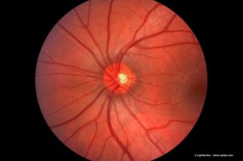
Evaluating both the genomic and proteomic biomarkers combined seems to be the most effective method to determine age-related macular degeneration (AMD) risk and the likelihood of progression to advanced AMD.
The greatest future challenge is the development of a blood test to identify individuals who will develop AMD before any signs of the disease become apparent, according to Dr Stephanie Hagstrom, PhD, who spoke during the retina subspeciality day at the annual meeting of the American Academy of Ophthalmology.
Complement components
Complement components already have been identified in drusen and a number of genetic studies have reported significant associations between AMD and DNA variants involved in the complementassociated pathway. Major breakthroughs in research during the past 5 years have shed a great deal of light on AMD and have affected the diagnosis, treatment and management of the disease, she pointed out.
Single nucleotide polymorphisms have been shown to be strongly associated with the risk of AMD and protection.
"Many of these factors are within the cellular components of the retina, such as the photoreceptor cells, the retinal pigment epithelium and the choriocapillaris," she added. "A number of these have also been identified in the acellular components, such as drusen."
Oxidative stress
One factor, oxidative stress, has been implicated in development of AMD because smoking has been shown to increase the risk of AMD and antioxidant therapy can slow the progression of AMD.
A direct link between oxidative stress and the development of AMD was found with the discovery by Dr Hagstrom's colleague, Dr John Crabb, PhD, that the concentration of carboxyethylpyrrole (CEP) adducts, an oxidative protein modification generated from docosahexaenoic acid (DHA), is elevated in Bruch's membrane and drusen. The combination of high exposure to environmental light and high oxygen tension from the choriocapillaris is the perfect setting for the generation of reactive oxygen capable of damaging DHA, a highly oxidizable fatty acid concentrated in the photoreceptor cells.
"Modifications in CEP are generated by covalent adduction of primary amino groups on DHA," Dr Hagstrom continued. "When generated, the oxidative fragments condense with amino groups on protein and become deposited in Bruch's membrane, the choroid, the retinal pigment epithelial cells, and drusen."
Such changes are thought to be antigenic and a source of inflammatory signal to initiate the pathology of AMD.
In her work to predict successfully the patients who are susceptible to developing AMD, she and her colleagues have genotyped more than 1400 patients for the AMD-risk polymorphisms and quantified the CEP biomarker levels in these patients.
Newsletter
Get the essential updates shaping the future of pharma manufacturing and compliance—subscribe today to Pharmaceutical Technology and never miss a breakthrough.


