Regaining presbyopic lens accommodation with FS laser cuts
Applying a specific number of FS-induced cuts to the crystalline lens could restore accommodation.
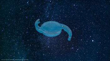
The treatment of presbyopia is one of the last frontiers in ophthalmology. Until now, the only treatment options that have been available to us have been the replacement of the natural lens with an intraocular lens (IOL) or the application of monovision concepts.
Presbyopia begins to be recognized around the age of 45, when the accommodative amplitude drops below 2 D. The crystalline lens becomes unable to form a spherical shape and it remains in the de or far-accommodated state whether the surrounding systems react or not. Nowadays, it is accepted that the ciliary body can still contract in presbyopic eyes, enough zonular fibres are present and the lens capsule remains elastic.1 Only the crystalline lens becomes denser and harder with age and all occurring forces are not strong enough to mold the lens.
Taking all of these factors into account, and following on from the research of a number of people, the Laser Zentrum Hannover in Germany has developed a new concept in presbyopia correction, which restores accommodation by improving crystalline lens flexibility. This is a theory that follows on from a study presented by Krueger and Myers ten years ago,2 where the researchers concluded that elasticity and deformation-ability could be changed by treatment with laser pulses.
It is well known that photodisruption with ultrashort laser pulses, during femtosecond (FS) LASIK procedures, offers an excellent and reliable tool for ophthalmic surgery. The cuts created by FS lasers show very few unwanted side effects and can therefore provide the necessary precision.
Having decided upon the ideal instrument to perform the small, precise cuts inside the crystalline lens, the procedure named lentotomy was created.
Lentotomy works on the premise that, if a laser wavelength in the near-infrared region is used, both the cornea and the crystalline lens are transparent for laser radiation. Thus, an FS laser can be focused inside the crystalline lens without opening the eyeball, generating a laser induced optical breakdown (LIOB) at the very focal point. While scanning the laser spot inside the lens, it is possible to disrupt tissue in a 3D-pattern. Thus, defined planes inside the lens tissue are created.
A previous study published by Krueger et al.,3 showed that, after in vivo cutting inside rabbit crystalline lens tissue, the lens stays clear for at least three months. In six treated rabbit eyes, laser-induced cataract did not occur and the three-month postoperative results also showed good transparency in these eyes. Thus, importantly, it has been shown that the application of small laser-induced cuts to the crystalline lens does not induce cataract formation.
Cutting the crystalline lens to restore accommodation
In order to regain crystalline lens elasticity and to increase crystalline lens deformation ability, respectively, we created gliding planes inside the lens tissue while leaving the lens capsule intact. These planes were generated by scanning the focus of the FS laser pulses in a defined pattern inside the crystalline lens.
In our studies,4 enucleated pig eye globes were fixated by a suction ring that fits to the curvature of the treated eye. For experiments with extracted lenses, only a much smaller fixation unit optimized in size was utilized. The ex vivo pig eyes were used within a few hours after enucleation. They were kept in saline solution to preserve the transparency of the cornea as well as the crystalline lens.
Newsletter
Get the essential updates shaping the future of pharma manufacturing and compliance—subscribe today to Pharmaceutical Technology and never miss a breakthrough.

