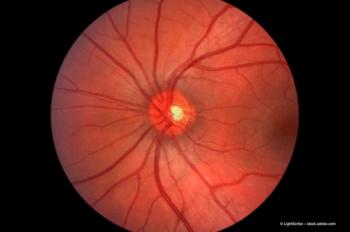
Raising the bar on retinal imaging
An electromagnetic deformable mirror reduces the impact of ocular aberrations on imaging for clearer results, according to Gisele Soubrane.
Key Points
Because of this ability to overcome the negative impact of aberrations on images, the Mirao 52-e electromagnetic deformable mirror (Imagine Eyes), used in conjunction with an adaptive optics flood illumination fundus camera (AOFIFC; INOVEO), can assist in more accurate diagnoses of ophthalmic diseases and conditions. This mirror and camera combination can also monitor the progression of these diseases and, ultimately, help in prescribing more targeted treatments that may have a positive impact on prognoses, according to Gisele Soubrane, MD.
"In combination with the electromagnetic mirror, the AOFIFC provides us with exquisitely sharp images of retinal microstructures," Dr Soubrane said. "The construction of the device is such that, after it has been altered by its course through the human optical system, the light entering the eye will be 'straightened-out', thereby producing much sharper images."
"The electromagnetic mirror has not yet been coupled with a clinical OCT instrument; however, we are very excited to see the images that this technology combination will produce, as we expect an even higher resolution than that provided by conventional OCT," she said. According to Dr Soubrane, the mirror raises the bar, not only in terms of the image quality, but also in terms of versatility and depth of field.
Imaging rods and cones
In addition to compensating for the eye's optical aberrations, the AOFIFC employs infrared light for imaging. This means that not only can the cone cells be imaged and their numbers counted accurately, but that, in the near future, their viability and metabolism could also be assessed.
"In some ocular diseases, the numbers of cones are decreased and, using this device, we are able to evaluate the status of the cells and see how well these cells are functioning in vivo," Dr Soubrane claimed. "The electromagnetic mirror enables the AOFIFC to offer us information down to the cellular level. Clinically, this is proving to be invaluable, assisting us in diagnosis and prognosis, as well as in determining therapies for certain ocular diseases and disorders. Disease progression can be tracked much more effectively with this device: this is crucial for a more favourable prognosis."
Because of their extremely small size, it is not currently possible to image rods. According to Dr Soubrane, however, the mirror may soon overcome even this.
"If another wavelength is used, and/or if the mirror is coupled with a magnifying device or system and adapted, we may in future be able to image these retinal cells better," she said.
How is this better than other devices?
Prior to the advent of the AOFIFC, investigative clinical retinal imaging was performed using fundus cameras, scanning laser ophthalmoscopes, or OCT; some of these had been previously enhanced with adaptive optics in restrictive laboratory settings. The mirror and the AOFIFC offer a different imaging perspective by substituting the high axial resolution achieved using OCT with more detail along the lateral axes.
"The two technologies work well together to offer a quality image of the retina, both in terms of resolution and depth, providing physicians with a refined diagnostic tool for identifying and managing ocular diseases," Dr Soubrane noted.
The mirror improves the resolution, contrast, and definition of retinal images, allowing for a six-fold improvement in their lateral resolution: from approximately 15–20 µm to 3 µm. This may help to understand those retinal diseases in which knowledge of the aetiology or the pathology remains elusive, and for which targeted treatment is scarce and hard to come by.
"The hope is that the AOFIFC, with its provision of more refined and detailed retinal images, will let us discover the aetiology of dysfunctional retinal cells, offering us a deeper understanding of their pathology," she said. "This may, in turn, allow us to delve into the origins of numerous ocular diseases and offer more effective treatments.
"The one downside of the AOFIFC's images is that the visual field available to stimulate the patient's fixation is only 20º, which is not large enough," Dr Soubrane stated. "This aspect of the imaging could use some improvement and fine-tuning. We need to experiment with other wavelengths in order to facilitate the evaluation of cells other than photoreceptors - including bipolar and ganglion cells - which may shed additional light on the influx of signals sent from the retina to the brain."
Application for glaucoma
It is now accepted that glaucoma is not simply an issue of increased IOP, and that analysis of the cellular damage around the optic nerve may offer some insight to the disease's aetiology.
"The cellular definition that the AOFIFC offers may make this evaluation possible," Dr Soubrane suggested. "The AOFIFC may be able to elucidate which cells are damaged, while providing information into the disease's progression. That information, in turn, may offer insight when choosing appropriate preventive treatments designed to spare these cells from disease and death," she continued.
"It is very important to have an in vivo cellular view, not only for accurate diagnosis of intraocular diseases but also for comprehending their progression. A deeper understanding of these pathologies at the cellular level will soon assist us in treating them much more effectively," Dr Soubrane concluded.
---------
Special ContributorGisele Soubrane, MD is a professor and chief of the Department of Ophthalmology at the Centre Hospitalier Intercommunal de Créteil, Créteil, France. She may be reached by E-mail:
Dr Soubrane is a member of Ophthalmology Times Europe's editorial advisory board.
Newsletter
Get the essential updates shaping the future of pharma manufacturing and compliance—subscribe today to Pharmaceutical Technology and never miss a breakthrough.


