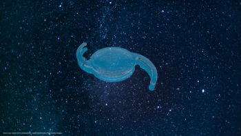
Pupil-expansion device addresses surgical challenges of small pupils
Prof. Ramin Khoramnia describes cases of posterior synechiae with very small pupils as well as his preferred insertion and removal technique.
One of the greatest challenges facing the ophthalmologist is performing surgery on eyes with small pupils. Small pupils add complexity and difficulty to many ophthalmic procedures. Devices which effectively open these troublesome pupils are important and useful. Pupil expansion devices are essential tools for cataract surgery in cases of non-dilating pupils.
One such device [I-Ring Pupil Expander, Beaver-Visitec International (BVI)] delivers a user-friendly intraoperative management of small pupils with its unique design and promising features. I have been using this device for years now-several procedures weekly-and find its simplicity and ease of use advantageous in my surgeries.
The pupil expansion device is designed to safely expand the iris tissue in order to provide the surgeon a 7-mm field of view intraoperatively. It is a single-use iris expander made of soft-yet-resilient polyurethane.
The ring engages the iris completely, expanding it evenly over 360°, creating a uniformly round opening of 6.3 mm in diameter.1
On the outside of the ring there are four corners pointing away from the central opening, creating four channels that hold the iris in place. Each corner contains a positioning hole for a Sinskey hook, separated from the channel in which the iris sits, to ensure that the Sinskey hook does not touch the iris during placement.1
The device comes packaged in a system with the inserter attached.
Indications for small pupils
I treat a lot of complex cataract cases, such as patients with small pupil and pseudoexfoliation syndrome, which require the use of a pupil expansion device.
I also use the device as a precaution in cases where I notice the pupil is not well dilating in the beginning as, in my experience, in these cases the pupil gets smaller during surgery. The device helps ensure that the pupil does not constrict.
Additionally, I have a lot of patients with posterior synechiae, for example, after uveitis, where after the initial lysis of the synechiae, the pupil is always very small and for me an indication to insert the device.
In some femtosecond-laser-assisted cataract cases, after the femtosecond laser procedure, the pupil becomes smaller and adrenalin injection is required to dilate it. If it does not completely open again, I will also use the device. Even in premium cases, I tend to use this device. It has become a standard procedure for us.
Insertion and placement of the device ensure a complete 360° engagement with the iris, providing a consistent pupil expansion without distortion.
My approach with posterior synechiae
In cases of posterior synechiae, with very small pupils, I find the device very useful. Firstly, I remove the synechiae, then I always inject more viscoelastic to try to dilate the pupil but usually it does not have any effect on these pupils. If pupil stretching also does not help, I don’t hesitate to use the device right away.
It is preferable and carries less risk to implant the device at the beginning of the procedure. The device can still be inserted once the capsulorhexis is complete; however, there is a risk of inserting it into the capsulorhexis. Therefore, increased vigilance is required to ensure the device is inserted in the right plane.
I favour the one-handed approach that accompanies the device insertion, and I like to use the second hand to keep the eye safe. With a local anaesthesia, and a patient who moves around a lot, I can stabilise the eye with the left hand and easily use the injector single-handedly. Once the device is in the eye, it is simple, with the Sinskey hook, to manipulate it and engage it into the iris.
When removing the device, I always place the inserter/remover in through the incision and then I grasp the distal edge of the device. As I disengage the device, it flips into the inserter/remover very quickly. The pupil looks perfect afterwards. Previously, iris hooks could leave damage to the iris, or other ocular structures. The device, however, does no damage to ocular structures as it is extremely gentle on the eye. This is a major advantage over traditional iris hooks and makes the pupil expander a go-to device in many complex small pupil cases.
Pupil expander features and benefits
A small pupil makes cataract surgery more difficult by:
- Limiting the size of the capsulorhexis.
- Making the nuclear disassembly more difficult.
- Increasing the risk of iris trauma.
- Reducing visualisation.3
The device can help alleviate these difficulties as it has a unique design structure and many beneficial elements, as described below:
> The polyurethane material reduces the risk of damage to the iris as it is very gentle on the tissue, yet its unique design firmly supports the entire pupillary margin.4
> Fixed channel height does not compress and pinch iris during insertion or removal.4
> There is relatively no learning curve: it’s very easy to use.
> Device insertion, engagement and removal are performed with great ease and avoid traumatic injury to the iris and surrounding tissue.
> The device allows for a single-handed procedure; this is particularly brilliant as it affords me a free second hand in which I can stabilise the eye, which is very advantageous in a difficult case.
> Green colour provides excellent contrast and visibility.
> Enhanced comfort: it is designed to remain planar in the anterior chamber, protecting corneal endothelium.4
> No additional incisions are required. Once the I-Ring is in the anterior chamber, it can be easily positioned by using the positioning holes. Less incisions are always favourable as there will be decreased risk of post-operative infection, inflammation and other complications.
> Aperture shape helps guide capsulorhexis.
> Minimal preparation for insertion by surgical team. It is simple to engage and extract and it is a single-handed fast technique.
> The device allows a uniform and round pupil expansion, unlike the diamond-shaped pupil that
is seen when iris hooks are used.
> The device does not lift the iris upward toward the incisions, which is the case with iris hooks. This is a great safety feature, decreasing the risk of damage to the iris
when entering the wound (e.g., with the phaco tip).
> Surgery is atraumatic and simple using the device.4
> Iris quickly returns to natural shape post-surgery.4
I like the device mainly because it is very easy to use. Insertion and removal of the device is very simple, intuitive and can be performed single handed. These features enable the surgeon to perform the most difficult cases with very small pupils with great ease.
Conclusion
The device creates a solution for intraoperative small pupil expansion, advancing the standard of small pupil management offering safety and reliability. The device is improving visualisation and is simple to implant and explant, making it a straightforward procedure.
The challenge posed by small pupils can now be effectively managed using the device and complex cases can have more successful outcomes thanks to this simple novel device.
Disclosures:
1. Priya Narang and Amar Agarwal, I-Ring expands pupil, enhances surgical field of view. Ocular Surgery News U.S. Edition, February 10, 2016. Available at:
2. www.bvimedical.com Available at:
3. Kent, C. Four New Ways to Manage Small Pupils, 5 NOVEMBER 2015. Available at:
4. www.bvimedical.com Available at:
Newsletter
Get the essential updates shaping the future of pharma manufacturing and compliance—subscribe today to Pharmaceutical Technology and never miss a breakthrough.


