
|Articles|April 1, 2006
Phaco complications: iatrogenic descemetorhexis
The corneal endothelium is able to function adequately, even after losing a large number of endothelial cells.
Advertisement
In a healthy, adult cornea, endothelial cell count is between 2,000 and 2,500 cells/mm2 with loss of corneal transparency occurring when this cell count falls below 500 cells/mm2 or when the corneal endothelium is damaged as a consequence of surgery, trauma or underlying disease, thus resulting in corneal oedema and loss of visual acuity. This situation often leads to a patient requiring a corneal transplant.
Here, Amar Agarwal describes a case report of unintentional iatrogenic descemetorhexis, arising during phacoemulsification, and the steps taken to overcome this complication.
A 65-year-old lady, who had previously undergone cataract surgery elsewhere, in the right eye, presented to us with complaints of decreased vision in the left eye, which had lasted for a period of six months. Upon examination of her records, a secondary anterior chamber intraocular lens (IOL) implantation in her right eye had been performed because of intraoperative posterior capsular rupture, which left the patient with intermittent iritis. She was also taking topical Betaxolol 0.5% twice daily for glaucoma.
Examination of the patient revealed the following attributes:
The first course of action
A decision was made to perform superior trabeculectomy in the patient's left eye with temporal clear corneal phacoemulsification and foldable IOL implantation.
During surgery, after the superficial scleral flap for the trabeculectomy, the clear corneal temporal incision for phaco was made. The side port incision was then made with a 26-gauge needle and viscoelastic was accidentally injected into the corneal stromal layers. As I did not recognize this complication at the time of surgery, a rhexis was performed in the space created by the viscoelastic substance. On attempting hydrodissection, I realized that the anterior lens capsule was intact and that I had inadvertently performed a "descemetorhexis". Subsequently, a capsulorhexis was performed and the remaining phacoemulsification and trabeculectomy were completed uneventfully.
How did the patient respond?
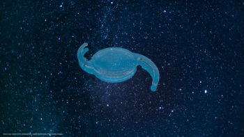
The patient was followed closely and one month later, we recorded a BCVA of 20/20p (6/6p), N6 in the left eye and a pachymetry of 560 and 556 microns in the right eye and left eye, respectively. By this time, the margins of descemetorhexis were clearly visible (Figure 1).
The second course of action
Newsletter
Get the essential updates shaping the future of pharma manufacturing and compliance—subscribe today to Pharmaceutical Technology and never miss a breakthrough.
Advertisement
Related Content
Advertisement


Latest CME


Advertisement
Advertisement