Oximetry reveals abnormal oxygen metabolism in exudative AMD
Professor Stef?nsson reveals the results of a comparative study demonstrating that observation of metabolic processes is possible enabling insight into the pathophysiology of disease.
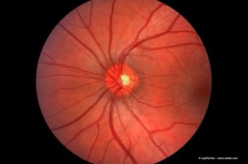
The design of the non-invasive retinal oximeter is based on a fundus camera. It simultaneously captures images of the retina at 600 nm and 570 nm and estimates retinal vessel oxygen saturation, then producing an image in which the degrees of oxygen saturation are indicated by different colours.
To establish whether the retina metabolises oxygen differently in an eye with AMD than in a healthy eye, the Oximetry Group in Reykjavik, including Asbjorg Geirsdottir, Sveinn Hakon Hardarson and professor Einar Stefánsson (University of Iceland, National University Hospital, Reykjavik, Iceland) compared the oximetry images of healthy eyes with those of eyes with exudative AMD.
When oxygen saturation in AMD patients is compared with healthy individuals a statistically significant difference is seen. While the venous oxygen saturation of healthy individuals decreases with age, in AMD patients the opposite pattern is seen.
He continued, "The preliminary results indicate that oxygen metabolism is different in healthy and AMD eyes."
Newsletter
Get the essential updates shaping the future of pharma manufacturing and compliance—subscribe today to Pharmaceutical Technology and never miss a breakthrough.


