|Articles|September 1, 2008
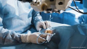
Lens tilt & decentration measurements made easy
Dr Frank Schaeffel & Dr Hakan Kaymak discuss a new device that measures tilt and decentration of the crystalline lens in both phakic eyes of healthy subjects.
Advertisement
Key Points
We know that tilt and decentration of the natural crystalline lens can significantly affect the optical quality of the foveal image; however, we know surprisingly little about how these factors are distributed in normal, healthy, phakic eyes, nor do we know the correlation of these variables between eyes. In recognition of this, one of us (Frank Schaeffel) has developed a simple, portable, easy-to-use, and partially automated measurement system to provide further insight.
What makes this system different?
To quantify the degree of decentration, the system uses image magnification, which is controlled by its low focus depth and by the modulation of the focus to produce sound of variable frequency.
How do we set it up?
No positional information was necessary for fixation targets, as the system incorporates a gaze tracker.
Measuring P3 initially caused several problems, in that it disappeared behind the iris when the fixation was a few degrees distant from the camera, and so P3 was difficult to find to begin with. During the initial trial of the system, it was found that, to provide the greatest contrast for optimal P3 visualization, an analogue charge-coupled device video camera must be used. P3 was also out of focus when the first (P1) and the fourth (P4) Purkinje images were at best focus (Figure 2); to obtain simultaneous measurements, the lens aperture had to be reduced to f/5.6 or f/8.
A green fixation LED was attached 2.53° below the IR LED. The LEDs were positioned 39 mm and 27.5 mm from the camera axis, equivalent to visual angles of 8.7° and 6.2°, respectively.
Advertisement
Related Content
Advertisement


Latest CME


Advertisement
Advertisement
Trending on Ophthalmology Times Europe
1
European Dry Eye Society Congress 2026: real-world evidence on Artelac Complete in dry eye disease
2
Demodex blepharitis: Closing the diagnostic gap to improve ocular surface health
3
Q&A: Uveitis risk rises as immune checkpoint inhibitor use expands
4
DEX implant shows benefit across RVO subgroups in real-world retrospective study
5