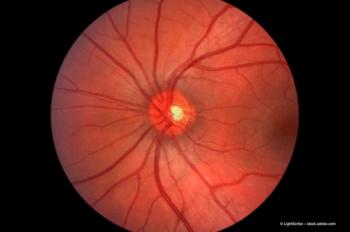
The IOL-VIP system: restoring sight to AMD patients
Dr José Luis Menezo looks at improving the visual acuity of patients with age-related macular degeneration (AMD) using an IOL-Vip system.
Key Points
The most commonly used low-vision device, the intraocular miniaturized telescope (IMT), confines the area of vision to a 20-degree angle at the central visual field and relies on the fellow eye to preserve the peripheral visual field. This, in my opinion, is not ideal. By contrast, the IOL-VIP (intraocular lens for visually impaired people) system, first presented in Florence in 2003,1 offers a better alternative. It does not compromise the peripheral visual field (leaving an area of approximately 80º) and can be implanted in both eyes, maintaining binocular vision.
A cross-discipline group of researchers from both the Medical University and the Physics University of Valencia2 presented a pilot study of the IOL VIP system in a series of 19 eyes at the 2008 ESCRS meeting in Berlin.3 The data gained from this pilot study were compared with the theoretical values obtained in a previous study.4
The cornea:anterior chamber lens distance and the anterior chamber lens:posterior chamber lens distance were both evaluated with high-frequency ultrasound biomicroscopy (UBM). Endothelial cell count was measured using a specular microscope preoperatively and 18 months postoperatively.
No complications were observed
Of the 19 patients enrolled in the pilot study, no cases of intraoperative or postoperative complications were detected, and no patients presented severe adverse effects. Four patients presented increased intraocular hypertension and a mild sectorial corneal oedema, controlled with topical timolol maleate. Only three patients developed posterior capsular opacification during the course of the study, which were treated with neodymium YAG laser. A single patient requested removal of the device after experiencing what was subjectively and confusedly described as a "strange situation" mixed with dizziness, but without diplopia. The refractive results in this patient were satisfactory; nevertheless, both lenses were removed and a standard monofocal lens was reimplanted.
Newsletter
Get the essential updates shaping the future of pharma manufacturing and compliance—subscribe today to Pharmaceutical Technology and never miss a breakthrough.


