|Articles|April 28, 2008
Intraocular stability of phakic IOL models may affect long-term results of IOL interaction with surrounding tissue
The intraocular position of different models of phakic IOLs show differences in space to the crystalline lens and the corneal endothelium, which may affect long-term results in terms of IOL interaction with surrounding tissue, according to a study published in the January 2008 issue of the Journal of Cataract & Refractive Surgery.
Advertisement
The intraocular position of different models of phakic IOLs show differences in space to the crystalline lens and the corneal endothelium, which may affect long-term results in terms of IOL interaction with surrounding tissue, according to a study published in the January 2008 issue of the Journal of Cataract & Refractive Surgery.
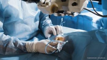
Thomas Kohnen, MD and team from the Johann Wolfgang Goethe-University, Germany, evaluated the postoperative intraocular positional stability of one rigid polymethylmethacrylate (PMMA) phakic IOL model and two foldable polysilicone-PMMA iris-fixated phakic IOL models.
One of three iris-fixated phakic IOL models (Artisan, Artiflex I, and Artiflex II; Ophtec BV) was implanted in 45 eyes of 26 patients with myopia or myopic astigmatism; each model was implanted in 15 eyes. The central distance between the phakic IOL and corneal endothelium and between the phakic IOL and anterior surface of the crystalline lens was evaluated using Scheimpflug photography six and 12 months after surgery.
The mean preoperative spherical equivalent was -9.32D±1.78 (SD) (range -6.5 to -13.5 D). The median distance from the central corneal endothelium to the anterior surface of the phakic IOL at six months and 12 months was 2.65 mm and 2.64 mm, respectively, in the Artisan group, 2.47 mm and 2.50 mm, respectively, in the Artiflex I group, and 2.48 mm and 2.52 mm, respectively, in the Artiflex II group. The median distance between the posterior surface of the phakic IOL and the anterior surface of the crystalline lens at six months and 12 months was 0.40 mm and 0.48 mm, respectively, in the Artisan group, 0.53 mm and 0.55 mm, respectively, in the Artiflex I group, and 0.68 mm and 0.66 mm, respectively, in the Artiflex II group. At 12 months, the distance between the phakic IOL and crystalline lens was statistically significantly greater in the Artiflex II group than in the Artisan group (p<0.01).
From these results, the researchers concluded that the intraocular position of rigid phakic IOLs and foldable silicone iris-supported phakic IOLs varied between the three phakic IOL models in space to the crystalline lens and the corneal endothelium, which has long-term implications in terms of IOL interaction with surrounding tissue.
Advertisement
Related Content
Advertisement


Latest CME


Advertisement
Advertisement