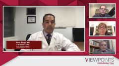
Imaging for diabetic retinopathy
The importance of imaging in a diabetes eye exam with OCT-A and widefield angiograms is examined.
Episodes in this series

Rishi Singh, MD: Dr Chous, you briefly mentioned the idea of imaging. What is the typical imaging pattern you’d end up choosing for some of the patients [such as] this?
A. Paul Chous, OD, MA, FAAO: Fundus photography is helpful, particularly widefield or ultra-widefield imaging….Some findings suggest that patients who have more peripheral diabetic retinopathy lesions are dramatically more likely to progress by 2 steps on the ETDRS [Early Treatment Diabetic Retinopathy Study] scale or ultimately develop proliferative disease. I’m also a fan of OCT [optical coherence tomography] imaging. I like to do OCT imaging every time I see a patient because I have a number of patients I’ve seen over the last 10 years who have subclinical diabetic macular edema [DME] that I simply didn’t detect with my normal fundus examination at the slit lamp.
Rishi Singh, MD: Dr Loewenstein, can you talk a bit about OCT-A [optical coherence tomography angiography] and the idea of using widefield angiograms in these patients? What have they led to as far as understanding these patients?
Anat Loewenstein, MD: Yes. It is more important to examine the posterior part and the midperiphery of these patients. I’m not against doing widefield imaging, but for diabetic patients most of the pathology is found in the midperiphery. Especially for screening purposes, widefield photography is fine, and I’m not doing widefield angiography on all the patients. I try to do angiography on any patient at baseline if they have some diabetic retinopathy. If they don’t have any diabetic retinopathy, I don’t do a fluorescein angiography at baseline even though they might have some nonperfusion and even though it might allude to the fact that they may have later progression on the diabetic retinopathy severity scale. At least for screening purposes, the most important things are the clinical examination and the OCT. I don’t trust myself [to not miss] mild cases of diabetic retinopathy.
What is a good clue is this: if a patient does not have diabetic retinopathy at all or if it’s only mild, the chances of them having diabetic macular edema are small. It is only if you have moderate NPDR [nonproliferative diabetic retinopathy] that you have higher chances to have macular edema. I am also doing OCT imaging on all my patients at baseline to make sure that I’m not missing something. This is what I am doing, but I’m not doing OCT-A imaging as a routine in every patient for baseline. If everything is fine, I’m not doing OCT-A imaging, mainly because of reimbursement issues. If every patient can have all the tests in the world, then I don’t mind doing OCT-A imaging, but the yield is not high in patients who don’t have clinical disease. I do OCT-A imaging if something is not clear to me during the follow-up of the patient or during the management while we are treating them.
Rishi Singh, MD: Your points are well taken, but I wonder about the value of OCT imaging in a patient with good vision. For a patient who has 20/20 vision or 20/25 vision, I’ve stopped getting OCT imaging for them because of some of the data we may talk about in our later sessions. We may talk about why the treatment indication isn’t there for that patient. I suspect it’s good for baseline testing, as you recommended, as well.
Anat Loewenstein, MD: Yes, I agree. For a patient with good vision acuity, according to the protocol of the DRCR [Diabetic Retinopathy Clinical Research] Network, you wouldn’t treat even if they have mild edema because it’s the same visual acuity results if you wait until they develop more severe edema. That’s only if you follow up with the patient. You’re absolutely right: it’s just for establishing a baseline.
A. Paul Chous, OD, MA, FAAO: I would add that the patients who do have mild DME have good visual acuity even though they’re not going to be treated. The data say that those patients are much more likely to progress on to DME that does affect their visual acuity. It’s a marker for the eye care professional to monitor that patient more closely.
Rishi Singh, MD: That’s a great point. Can you talk a bit about widefield angiography, Anat? OCT imaging is non–dye-based, but you’re talking about dye-based angiography. What have you found from the widefield imaging studies that you’ve done in patients?
Anat Loewenstein, MD: I don’t do widefield angiography as a routine. I don’t think it changes the way I will treat a specific patient. What is right about it was said before: if you have severe nonperfusion in the periphery, you might want to follow up on the patients more closely. However, it will not change your treatment at this point at all, so it is not in my routine to perform it. According to older studies, [such as] the ETDRS and other studies, nonperfusion in the periphery is not an indication for treatment. It is only the case if you have neovascularization. I also am not in the European group that believes that you improve the macular edema if you do laser therapy to areas of nonperfusion in the periphery. There is enough data to show that it does not do that, so to me, it is not routine to do widefield angiography. If I don’t understand something, then I might do it.
A panel of experts in ophthalmology and optometry review the diagnosis and treatment of diabetic eye disease including emerging agents in the field.
Newsletter
Get the essential updates shaping the future of pharma manufacturing and compliance—subscribe today to Pharmaceutical Technology and never miss a breakthrough.


