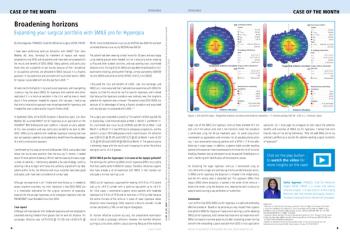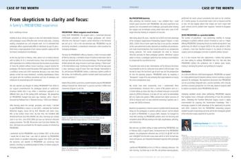
From Optical to Digital Precision: Advancing Retinal Surgery with the ARTEVO 850 Microscope

Surgical microscopes have undergone significant changes over the past decade, moving from conventional optics toward digital platforms that redefine the boundaries of surgical visualization. My own transition from the ARTEVO® 800 (Carl Zeiss Meditec AG, Jena, Germany) to the ARTEVO® 850 microscope has provided a firsthand perspective on how these advancements translate into real-world clinical benefit—particularly in the management of vitreoretinal surgery.
Surgical microscopes have undergone significant changes over the past decade, moving from conventional optics toward digital platforms that redefine the boundaries of surgical visualization. My own transition from the ARTEVO® 800 (Carl Zeiss Meditec AG, Jena, Germany) to the ARTEVO® 850 microscope has provided a firsthand perspective on how these advancements translate into real-world clinical benefit—particularly in the management of vitreoretinal surgery.
From ARTEVO 800 to ARTEVO 850: A Paradigm Shift in Visualization
We entered the era of digital ophthalmic visualization with the ZEISS ARTEVO platform. The ARTEVO 850 has not simply improved upon its predecessor, the ARTEVO 800. Rather, it has refined how surgeons interact with the ocular anatomy at a fundamental level. Nowhere is this transformation more evident than in three domains critical to microsurgical finesse: illumination, color management, and depth of field.
The ARTEVO 850 redefines surgical lighting with a pure RGB LED system and digital color assistant (DCA). Equipped with a mix of red, green, and blue LEDs and with its DCA technology, the ARTEVO 850 allows surgeons to adjust light color temperature from warm to cold (3000K to 6000K) in increments of 250K. In my opinion, this degree of tunability is especially meaningful in retinal surgery where ambient contrast and hue can influence dissection decisions. Moreover, the uniform illumination of the ARTEVO 850 ensures that field brightness is preserved even at lower intensity levels, maintaining visual fidelity while minimizing stress on the retina. This enhancement reflects an ongoing trend in digital visualization aiming to not only increase clarity, but to also make illumination biologically safer and visually more intelligent.
Surgical success often depends on detecting minute variations in tissue tone, staining behavior, or hemorrhagic states. While the ARTEVO 800 excels in delivering natural color reproduction, which is particularly useful when visualizing unstained membranes or differentiating neurosensory retina from underlying layers, the ARTEVO 850 builds on this foundation with the DCA that moves beyond faithful representation into the realm of color optimization. Surgeons can now digitally emphasize specific hues or modulate contrast based on the surgical step. Subtle epiretinal membranes (ERMs) can be digitally enhanced during peeling, and subretinal fluid pockets can be more easily distinguished without reliance on traditional stains or contrast agents. This level of color intelligence is not merely aesthetic—it is clinically functional, guiding the surgeon through complex anatomical differentiation in real time.
Another key element of digital visualization is how well the system handles depth of field (DoF)—how much of the surgical volume appears in crisp focus at any one moment. The ARTEVO 800 already offered a substantial advance here, leveraging digital optics to create a naturally extended DoF, reducing the need for constant refocusing and enhancing fluidity of movement during retinal or anterior segment surgery. The ARTEVO 850 introduces surgeon-controlled dynamic modulation of depth known as Smart Depth of Field. This new function allows real-time DoF adjustment depending on the demands of the surgical step. When precision at a single plane is needed, such as during internal limiting membrane (ILM) peeling, narrower DoF can enhance contrast. Conversely, during fluid-air exchange or subretinal injections, an expanded DoF (boost by 60% as compared to ARTEVO 800) allows visualization of the entire relevant anatomical span without optical compromise. And critically, this is achieved without increasing light intensity, preserving retinal safety while maintaining visual sharpness.
Clinical Experience: Vitrectomy and Macular Surgery
When performing routine core vitrectomy with the ARTEVO 850, the microscope’s “vitrectomy blue” filter refines visualization of the central vitreous compared to standard RGB modes, especially during peripheral shaving under scleral indentation (Figures 1 and 2). In my opinion, the contrast enhancement offered by this filter allows for safer and more efficient vitreous removal, potentially reducing iatrogenic traction.
During ERM peeling, I find that the “peeling blue” filter consistently improves intraoperative visibility of the membrane interface, eliminating the need for dye in some cases. Despite the technological advancements available on the ARTEVO 850, however, certain situations continue to present visualization challenges. For example, posterior staphyloma, macular schisis, and retinal pigment epithelium atrophy in highly myopic eyes often limit clarity even with spectral filtering. In these cases, combining the ARTEVO 850 filter technology with vital dyes such as Membrane Blue Dual proves most effective. The filter reduces background interference and minimizes glare, while the dye selectively stains the target tissue, allowing for more complete removal. The improved visualization using the blue filter allows for a single staining pass in most cases, decreasing procedure time and dye exposure.
Nevertheless, use of the filters alone has limited benefit in eyes with significant structural distortion. This highlights the continued need for tailored strategies in complex surgeries, including careful dye application protocols to avoid under- or over-staining. This is an area where standardization across practices is still needed.
The “peeling blue” filter has also proved advantageous in macular hole surgery. I find it enhances ILM contrast and delineation even in the absence of dye, although vital dye use remains preferred in most cases. The green filter serves as a useful adjunct in select situations, particularly where ILM pigmentation or retinal layering make visualization more challenging. The use of the Digital Color Assistant allows further optimization, reducing dye reliance and improving the visualization of fine structural changes at the fovea.
Intraoperative videos from a routine case of ERM peeling and in a complicated case involving an exudative, tractional ERM in an eye with a history of retinal vein occlusion highlight how the advanced features of the ARTEVO 850 enable surgical efficiency, precision, and safety (Figures 3 and 4). These cases also showcase the integrated optical coherence tomography that is available as an optional add-on feature for the ARTEVO 850.
Conclusion
The shift from the ARTEVO 800 to the ARTEVO 850 reflects more than just a technological upgrade—it represents a conceptual evolution in how we approach retinal surgery. Along with its ergonomic heads-up display, I find that the combination of high-resolution digital imaging, spectral filters, and intelligent depth modulation with the ARTEVO 850 provides an unparalleled surgical environment. Its innovations have translated to improved visualization during every critical step of macular surgery, from vitrectomy to membrane peeling and ILM dissection. While challenges persist, particularly in complex myopic anatomy, the ARTEVO 850 has expanded the possibility to see better and operate more precisely—hallmarks of progress in the digital era of ophthalmic surgery.
Reference
Mastropasqua R, Quarta A, Ruggeri ML, Mastropasqua L. Enhancing Precision and Clarity with New Digital Color Assistant in 3D Heads-Up Vitreoretinal Surgery. Ophthalmol Ther. 2025;14(4):805-814. doi:10.1007/s40123-025-01106-1
Dr Mastropasqua is a Full Professor of Ophthalmology at G. D’Annunzio University of Chieti-Pescara, Italy, and Director of the Surgical Visual Rehabilitation Department, ASL Lanciano-Vasto-Chieti, Italy.
Newsletter
Get the essential updates shaping the future of pharma manufacturing and compliance—subscribe today to Pharmaceutical Technology and never miss a breakthrough.


