|Articles|February 1, 2009
Flap adhesion under the spotlight
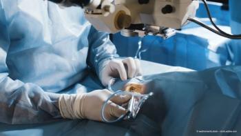
Professor Michael Knorz discusses his experiments on the adhesion strength of flaps, comparing the femtosecond with the microkeratome.
Advertisement
Key Points
Although there has been much discussion over whether mechanical microkeratomes or femtosecond lasers are better for performing refractive surgery in terms of both visual outcomes and safety, there remains one key metric that is essential for measuring the success of the procedure: flap stability.
Flaps created with femtosecond lasers have previously been shown to provide greater stability when compared with microkeratome flap creation.1 Michael C. Knorz, MD had noticed in his clinic during LASIK retreatments that femtosecond flaps appeared more difficult to lift than microkeratome flaps, and so he conducted a study to evaluate the effects of the cutting tool (as well as cut angle and, with the femtosecond, laser energy) on flap stability. Professor Knorz used AMO's IntraLase iFS femtosecond laser and compared the results with those obtained using Ziemer's Amadeus II microkeratome.
"We conducted these tests on rabbit eyes. The 14 rabbits were divided into four groups, and I conducted all the surgeries personally," said Professor Knorz. "We know that surface quality, flap thickness and shape, and corneal biomechanics and aberrations can influence surgical outcomes, but we concentrated on flap stability, and which instrument created the more stable flaps."
The microkeratome group included five eyes, and Professor Knorz created the nasal hinge using a 9 mm suction ring and a 140 µm head. The remaining three groups were operated on with different configurations of the IntraLase iFS femtosecond laser.
"In each of the femtosecond groups, we created 120 µm flaps with a diameter of 9 mm by using a repetition rate of 150 kHz. Group Two used a normal energy (0.8 µJ) side-cut on four eyes; Group Three used a high energy (1.6 µJ) side-cut on four eyes, and the final group of four eyes was operated on using an inverted side-cut, with an angle of 140° as opposed to 70°," explained Professor Knorz.
All the eyes with normal flaps were clear by day six, and this state maintained until the end of the study period (day 75). Of the corneas with amputated flaps that developed haze, most had cleared by day 75.
Postop flap excision: testing adhesion strength
For the first 15 days postoperatively, all eyes were treated three times daily with gentamicin sulphate 0.3% ophthalmic solution and dexamethasone
1 mg/mL ophthalmic solution. At day 75, the rabbits were euthanized and their corneal epithelia were excised with a hockey knife.
"After the epithelium was removed with a hockey knife, we used cynoacrylate ester to glue the flap surface to a plano-concave acrylic lens, which had a 10 mm radius curve and was attached to an aluminium holder. Both the acrylic lens and the holder were custom-made by AMO, and we were very careful to ensure that we glued just the flap and left the peripheral cornea untouched," clarified Professor Knorz. "Once the glue had been applied, we applied moderate pressure manually for six minutes while the glue dried."
A digital force gauge, model 475040 (Extech Instruments) was the tension meter used to measure adhesion strength. This was achieved by attaching the tension meter to the aluminium holder with a rope and pulling - at an angle of approximately 60° towards the hinge - until the flap dehisced.
Advertisement
Related Content
Advertisement


Latest CME


Advertisement
Advertisement
Trending on Ophthalmology Times Europe
1
Beyond anti-VEGF: The next wave in retinal therapy
2
Demodex blepharitis: Closing the diagnostic gap to improve ocular surface health
3
Trehalose-hyaluronate combination aids tear film after eyelid surgery
4
ZEISS reports early implantations of AT LUCIA Toric 721P IOL in Europe
5