Femtosecond gets thumbs up from Pallikaris
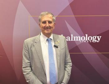
Femtosecond lasers provide a safe and effective way to create corneal flaps and tunnels, explained Professor Ionannis Pallikaris.
Femtosecond lasers provide a safe and effective way to create corneal flaps and tunnels, explained Professor Ionannis Pallikaris.
Using a femtosecond laser decreases the energy necessary to incise tissues, and it causes minimal thermal damage to surrounding tissues. He said another advantage of the laser was that it was an accurate, safe and predictable procedure with a high repeatability rate.
"It is a non contact process and there is a continuous line of new applications emerging," he said.
For flaps, the femtosecond laser creates thousands of laser pulse bubbles connected together in a raster pattern to separate the flap from the cornea. "LASIK flap creation with the femtosecond laser is the most common procedure and there are more than 1 million procedures performed each year," he said.
He said the advantages of a femtosecond laser, compared with a microkeratome, included accuracy. "You never get a thick-thin, unpredictable flap. Moreover flap thickness I irrelevant to preoperative corneal curvature, thickness, translation speed and IOP. And the risk of corneal ectasia are also minimized," he said, adding that the laser produced clear flaps, with no debris, no sterilization and minimized risk of infection.
To illustrate the issue with flap thickness, Dr Pallikaris showed a slide illustrating a microkeratome flap. In tests, applanation microkeratomes created very uneven flaps, with thick peripheries and thin centres, though he noted in passing that the indentation microkeratome was an improvement.
He said the femtosecond flap provided a better flap architecture and there were minimal induced aberrations, less corneal flattening and better hyperopic treatments.
He said there were some disadvantages compared with microkeratomes, saying the laser is bigger and describing it as a macrokeratome. It is also expensive and it created new complications, such as vertical gas breakthrough and reported cases of transient light sensitivity, for example.
Moving onto tunnels and intracorneal ring segments (Intacs), Dr Pallikaris said that tunnels were created for keratoconus and iatrogenic corneal ectasia. For these procedures the femtosecond laser enhances safety by providing greater consistency of depth and uniformity along the channel.
Dr Pallikaris showed examples of femtosecond-assisted biopsy and femtosecond assisted lamellar keratoplasty. And he finished his examination of femtosecond procedures with some slides detailing new information on femtosecond assisted posterior lamellar keratoplasty.
"In cases with isolated posterior corneal pathology, it is better to transplant on the diseased posterior cornea and leave intact the normal anterior layers," he said, explaining the logic behind FS-PLK.
He said femtosecond laser-assisted PLK has been performed in human eye bank eyes by HK Soong and colleagues and they showed shallow concentric ridges. In study with in vivo rabbit models, clinical appearance, consistent with corneal ectasia was noted in three out of 12 eyes.
Dr Pallikaris said that the FS-PLK would allow different shaped cuts to be used for different problems. For example, the zigzag shape provides a smooth transition between host and donor tissue and allows for a hermetic wound seal.
The top hat shaped incision allows for the transplantation of large endothelial surfaces, while the mushroom shaped incision preserves more host endothelium than the traditional trephine approach.
He concluded that new applications for the femtosecond techniques will continue to emerge.
Newsletter
Get the essential updates shaping the future of pharma manufacturing and compliance—subscribe today to Pharmaceutical Technology and never miss a breakthrough.


