Femtosecond benefits in corneal procedures
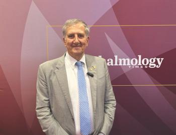
Dr Rudi Nujits, presented new research on femtosecond laser assisted Descemet Stripping Endothelial Keratoplasty (FS-DSEK) at a session on the femtosecond laser and therapeutic corneal surgery.
Dr Rudi Nujits, presented new research on femtosecond laser assisted Descemet Stripping Endothelial Keratoplasty (FS-DSEK) at a session on the femtosecond laser and therapeutic corneal surgery.
He said full-thickness penetrating keratoplasty has been the preferred corneal transplantation technique since the first description in 1905 by Eduard Zirm. "Although penetrating keratoplasty nowadays generally results in clear corneal grafts with a graft survival up to 72% at five years, the procedure is frequently complicated by refractive imperfections and wound healing problems," he said.
In 2005, 100 years after Zirm, the femtosecond laser was introduced to corneal transplantation surgery. "It is now possible to use the FS laser for combinations of lamellar and perforating cut patterns and for posterior lamellar keratoplasty," he said.
The idea behind the use of the FS laser is that it provides a precisely shaped corneal button that would be impossible to achieve manually. "This is an exciting technology with limitless potential in cutting constructions like top hat, inverted mushroom, or zig-zag shaped patterns for lamellar and penetrating keratoplasty.
He said several endothelial keratoplasty (EK) procedures, such as PLK, femto-PLAK, DLEK, DSEK, DSAEK, FS-DSEK, and DMEK, allow for selective replacement of the diseased endothelial layer retaining the healthy recipient anterior corneal stroma. EK techniques result in a rapid visual rehabilitation and minimal change in corneal astigmatism.
In a previous study, he said, Dr Nujits and colleagues showed that it is feasible to prepare a posterior lamellar disk (PLD).
In a recent prospective randomized multicentre study, as a part of the Dutch Lamellar Corneal Transplantation Study (DLCTS) they evaluated FS-DSEK (n=36) versus PKP (n=40). Excluding eyes with retinal disease, the 12-month logMAR BSCVA was 0.51 in the FS-DSEK eyes and 0.35 in PKP eyes (p
"The average BSCVA in our series appears to be lower as compared with a recent DSAEK series. Possible explanations for this finding are the quality of the interface at the stromal side of the PLD as prepared by the FS laser and an increase in interface haze after FS-DSEK due to activation of keratocytes, which results in more scatter," he noted.
He said the spherical equivalent was more hyperopic in the FS-DSEK group postoperatively, which was caused by the fact that the button is thicker peripherally and thinner in the centre.
Topographic astigmatism was 1.58 ± 1.2 D in FS-DSEK eyes versus 3.67 ± 1.8 D in PKP eyes (p
However, endothelial cell viability was not affected by the preparation of the posterior lamellar disc with the femtosecond laser, because preparation of the PLD with the FS laser resulted in endothelial cell damage of only 3.4 ± 3.5%.
"However, we should be concerned with preventing causes of cell loss in general," he said. "Drawbacks of current posterior lamellar procedures are extensive endothelial cell loss and a slightly lower BCVA," Dr Nujits said. "Technological improvements are needed to overcome these disadvantages and randomized clinical trials should carefully define the efficacy of this new technology."
Newsletter
Get the essential updates shaping the future of pharma manufacturing and compliance—subscribe today to Pharmaceutical Technology and never miss a breakthrough.


