
|Articles|January 1, 2011
FAF technology encouraging in evaluating AMD, study finds
Results of a recent clinical study provide promising evidence of the value of ultra-widefield fundus autofluorescence (FAF) imaging in the evaluation of age-related macular degernation (AMD)
Advertisement
Results of a recent clinical study provide promising evidence of the value of ultra-widefield fundus autofluorescence (FAF) imaging in the evaluation of age-related macular degeneration (AMD), according to lead investigator Dr SriniVas Sadda, MD, who presented the findings of the study at the annual meeting of the American Society of Retina Specialists.
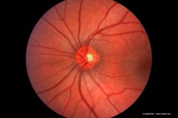
In a study using FAF imaging technology (200Tx [formerly the P200MAAF multi-wavelength scanning laser ophthalmoscope], Optos), peripheral FAF abnormalities were found in 77% of eyes in the series, especially in those with retinal degenerations, inflammation, and AMD.
Using the images, 174 eyes showed peripheral changes outside the central pole area of the retina, the area viewed using standard narrow-field, 30° field of view imaging technology. The findings demonstrated that peripheral FAF abnormalities often correlate with areas of retinal pigment epithelium disturbance. Also, when correlation occurs, the abnormalities often appear more striking on FAF imaging than on the pseudocolor images due to better contrast, according to Dr Sadda, associate Professor of Ophthalmology and director at the Medical Retina Unit, Ophthalmic Imaging Unit, and the Doheny Image Reading Center at the Doheny Eye Institute, University of Southern California, Los Angeles.
"The significance of these findings still needs to be defined and requires further study," Dr Sadda said. "However, the evaluation of peripheral FAF abnormalities may aid in diagnosis and provide new insights into disease pathogenesis. This study follows the recently published results from the Reykjavik/Moorfields study and adds to the growing body of evidence of the importance of studying the periphery of the retina in the diagnosis and management of AMD."
The retrospective analysis reviewed the records of all patients referred to the Doheny Eye Institute imaging unit for FAF imaging who had widefield images (200CAF or 200Tx, Optos) obtained over a 6-month period. The research team collected widefield and non-widefield FAF images obtained at the same visit as well as demographic data, including disease diagnosis.
All widefield images were evaluated for the presence of FAF abnormalities, defined as increased or decreased, beyond the central 30°. The frequency of abnormalities was computed and FAF abnormalities within disease categories were scrutinized for characteristic patterns.
The analysis included 225 eyes of 122 patients with ultra-widefield imaging. In all, 174 eyes (77%) had abnormal peripheral FAF studies. This included 100% of the eyes with a diagnosis of ocular tumors (n = 6) or retinal degenerations (n = 31) and 90% of those with inflammatory or infectious retinal diseases diagnosed (n = 54). The analysis also revealed that 76% of eyes with AMD (n = 34) and 63% of those with miscellaneous eye diseases (n = 45) had abnormal peripheral FAF imaging, as did 36% (n = 4) of the eyes with central serous chorioretinopathy.
Newsletter
Get the essential updates shaping the future of pharma manufacturing and compliance—subscribe today to Pharmaceutical Technology and never miss a breakthrough.
Advertisement
Related Content
Advertisement


Latest CME


Advertisement
Advertisement