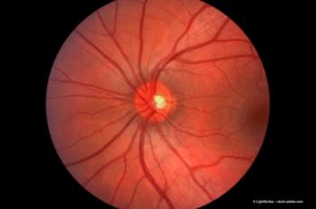
Evaluating Patients for a Retinal Disorder
Types of tests and criteria that can be used to diagnose conditions such as age-related macular degeneration, diabetic macular edema, and inherited retinal disease.
Episodes in this series

Albert J. Augustin, MD: Let us speak a little bit about a diagnosis process. Dr. Korobelnik, we would like to focus a little bit on fluorescein ICG [indocyanine green angiography] and OCT [optical coherence tomography]. Perhaps you can say a few words on multimodal imaging?
Jean-Franҫois Korobelnik, MD: I believe multimodal imaging is currently very important. What we do for macular disease is a fundus photography plus OCT. With that combination, we usually make the diagnosis in many cases. From time to time, we may use fundus autofluorescence. Fluorescein angiography for macular disease is less often used nowadays, especially since we have OCTA [optical coherence tomography angiography]. However, wide field fluorescein angiography is very interesting. We use FA [fluorescein angiography] wide field in several diseases, especially diabetic retinopathy, to evaluate the situation or to follow the treatment with combination with wide field color imaging.
Albert J. Augustin, MD: Thank you very much. Dr. Moosajee, is there a difference when we focus on hereditary diseases?
Mariya Moosajee, MBBS, BSc, PhD, FRCOphth: The diagnostic process for IRDs [inherited retinal disease] starts with a history; you can tease out whether this is a rod or cone dominant disorder by the symptoms. If it is rod predominant, then the patients will complain of night blindness, visual field loss, and then later on central vision loss. With cone predominant disorders, such as macular dystrophy or Stargardt disease, the central vision will be affected first; problems with near and distant vision: seeing details of faces, numbers on the bus, and then color and contrast and even photosensitivity. Taking an accurate family history is critical; asking about consanguinity. Then, in terms of investigations OCT, auto fluorescence, visual fields, and ERGs [electroretinograms] are extremely good for diagnostics. These are not so good for monitoring in routine clinics, but for me, again, it is all about genetic testing. That is the key to understanding the causative gene, and then access to potential treatments and trials.
Transcript edited for clarity.
Newsletter
Get the essential updates shaping the future of pharma manufacturing and compliance—subscribe today to Pharmaceutical Technology and never miss a breakthrough.


