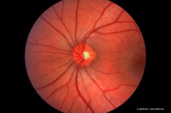
Diabetic macular oedema after phacoemulsification
Foveal thickness and macular volume decrease after phaco in eyes with diabetic macular oedema, with changes even more pronounced in patients with diabetic retinopathy.
Key Points
Several studies have determined that DME deteriorates after phaco, although there is, as yet, no consensus regarding either the degree or the prevalence of this decline. Likewise, it has been shown that phacoemulsification is associated with progression of DR in up to one third of cases.
To quantify the specific interactions between phaco and DME and DR, Dr Ken Hayashi and colleagues conducted a study examining post-phaco changes to DME in eyes with and without DR.
"We recruited 70 DME patients scheduled to undergo cataract extraction with intraocular lens implantation - 35 with diabetic retinopathy and 35 without - between May 2004 and July 2005," explained Dr Hayashi. "The number of eyes we examined was small, but we believe that the distribution of the stage of DR in our study accurately reflected the distribution within the population."
Patients were excluded from the study on the basis of the presence of cataract not caused by age or diabetes, a history of ocular inflammation or surgery, any macula or optic nerve pathology not caused by diabetes, and planned extracapsular extraction. The mean age of the patients included in the study was 67.9±7.0 years; patients in both groups were similar in terms of systemic diseases (hypertension and nephropathy) and ratio of right to left eyes, although the DR group had significantly increased percentages of haemoglobin A1C and duration of diabetes when compared with the no-DR group.
"To ensure consistency, I performed all the surgeries personally, using the same technique, and all patients were implanted with a hydrophobic acrylic IOL - either the MA60AC or the MA50BM models," Dr Hayashi noted.
The surgical technique
After making a 3.5 mm incision in the sclera, Dr Hayashi used a bent needle to achieve a continuous curvilinear capsulorhexis, intended to be slightly smaller than the optic diameter and to overlie the IOL optic circumferentially.
"I then performed nucleus and cortical aspiration, after completing the hydrodissection," Dr Hayashi noted. "To prepare for IOL implantation, I increased the wound size of 4.1 mm with a keratome, and inflated the lens capsule with Healon," AMO's sodium hyaluronate 1% product.
After preparing the capsular bag as described, Dr Hayashi inserted the IOL and evacuated all the viscoelastic material.
"All the surgeries went smoothly: there were no unexpected events or difficulties with IOL implantation," Dr Hayashi commented. "There were no significant differences between the two groups in the grade of the nucleus, ultrasound time, ultrasound power or infusion volume."
Newsletter
Get the essential updates shaping the future of pharma manufacturing and compliance—subscribe today to Pharmaceutical Technology and never miss a breakthrough.


