Amniotic membrane could move the needle outside of the operating room
The tissue serves as a testing ground for outpatient ocular therapies


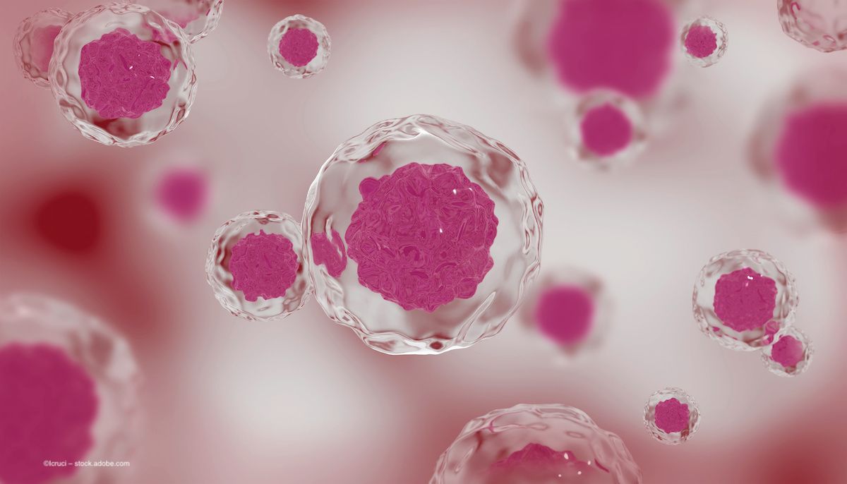
Amniotic tissue has an established history in eye care, but new applications could extend its efficacy from the surgical suite to outpatient and preventative therapies. Image credit: ©Icruci – stock.adobe.com
Infrastructure deficiencies have been a longstanding barrier to the use of cell and tissue therapies in an ophthalmic setting. New approaches to preservation, distribution and application have expanded the benefits of novel therapies to more patients. Along with regenerative approaches to grafts, such as stem cell products, amniotic membrane has also been recognised for its ocular applications. To alleviate the logistic burden around preparing, storing and reactivating amniotic tissue, Andrew Hopkinson, PhD, of the University of Nottingham in the UK, developed a novel method for processing and preserving the membrane; that technology was patented by the University of Nottingham and licensed to NuVision Biotherapies, where Hopkinson is now chief scientific officer.
The vacuum-dry preservation process resulted in a human amniotic membrane-derived dry matrix (Omnigen, NuVision Biotherapies) that can be applied to the eye as a standalone patch, as a graft or as part of a bandage contact lens (BCL) which holds the tissue to the eye (OmniLenz). When used in a surgical setting, this amniotic membrane has proven effective for patients undergoing epithelial redirection to manage limbal stem cell deficiency (LSCD) following stem cell transplant.1 Investigators Dua et al assessed patients for LSCD following chemical eye injury, congenital aniridia-related keratopathy and other ocular surface injuries. Vacuum-dried amnion and fibrin glue were used in a modified conjunctival epithelial redirection technique. Amniotic membrane has also taken the place of conjunctival autograft in pterygium management surgery, resulting in a sutureless procedure with shorter operation time.2
The lens-mounted membrane was well tolerated by patients in recovery from acute chemical eye injuries.3 In that study, 21 patients (23 eyes) with acute chemical eye injury received first aid followed by application of amniotic tissue within 2 days. Some patients underwent repeated application of the lens (five eyes) and the size of epithelial defect was reduced after a second application. Within 1 month, complete epithelial healing was achieved in all eyes.
Amniotic membrane has unique qualities that lend it to several effective ocular applications and make it a strong candidate for innovative preservation, storage and usage methods. In conversation with Ophthalmology Times Europe, Andy Hill, CEO of NuVision, discussed qualities of the amniotic tissue product that are specifically designed to target the logistic and infrastructure challenges considered endemic to these therapies. Hill explained that the membrane is room-temperature stable and can be stored at any temperature between 2° C and 25° C. In addition, rehydration is nearly instantaneous; in the case of the amnion BCL, the tissue is rehydrated by the solution the contact lens is stored in. These qualities make it available at the point of use with little preparation time required.
The ease of access, combined with the lack of reliance on sutures, indicate that amniotic membrane has robust potential outside the surgical suite. While the vacuum-preserved amnion was developed with a surgical setting in mind, Hill estimates that “approximately 80% of the product’s usage happens in the outpatient department or a physician’s office.” As such, the company is exploring new applications for the material to meet a range of patient needs. Currently, Hill said, investigators are concluding a randomised control trial on applications of amniotic tissue for dry eye disease at Aston University in Birmingham, UK. Data from that trial are not publicly available yet. However, according to an infographic from NuVision detailing initial characteristics of the prospective study,4 the trial as designed includes 35 patients (70 eyes) with an ocular surface disease index (OSDI) between 25 and 80. The trial includes patients with at least one of these clinical criteria: corneal staining (five or more punctate spots), conjunctival staining (nine or more punctate spots) and noninvasive Keratograph tear breakup time of 8 seconds or less.
The proprietary Omnigen and OmniLenz materials are currently in use at 130 hospitals in the United Kingdom, including Moorfields Eye Hospital, and an additional 50 private hospitals, Hill said. However, expanding throughout the UK, into Europe and beyond will take time and effort. Hill noted that while the tissue is shelf stable for up to 60 weeks, to store human tissue for longer than 48 hours, practices must have the relevant license from the Human Tissue Authority. Larger hospitals and health care facilities would likely have the proper licensing to keep amnion on hand, but this could present a burden of use for smaller private practices. Currently, NuVision is working to establish a European manufacturing base for tissue derived in Europe. As production expands into the European continent, the company may face policies that continue to change with the industry; the European Commission introduced new regulations on quality and safety standards for substances of human origin in December 2023.
Hill said that he hopes to expand the company's offerings into the United States as well, and to widen the applications for amnion within the ophthalmic and optometric markets. The company is leading exploratory research into amniotic tissue’s capabilities as a delivery system for medications, to reduce the patient burden of topical applications such as eye drops, and as a pre-surgery stabilisation technique for patients undergoing refractive surgery. In the posterior segment, prior research has indicated that amniotic tissue is a strong candidate for plugging macular holes.5 Amniotic plug transplants using traditionally preserved amniotic tissue have been successful, and use of a vacuum-preserved amnion graft could make the procedure accessible to more patients.
References
1. Dua HS, Ting DSJ, AlSaadi A, Said DG. Management of limbal stem cell deficiency by amnion-assisted conjunctival epithelial redirection using vacuum-dried amniotic membrane and fibrin glue. Br J Ophthalmol. 2023;107(3):342-348. doi:10.1136/bjophthalmol-2020-318496
2. Xanthopoulou PT, Elanwar M, Alzyadi M, Lavaris A, Kopsachilis N. Α novel sutureless pterygium excision surgery using human-derived dehydrated amniotic membrane. Cureus. 2022;14(4):e23839. Published 2022 Apr 5. doi:10.7759/cureus.23839
3. Lotfy NM, Al Rashidi S, Hagras SM. Clinical outcomes of vacuum-dehydrated amniotic membrane (Omnigen) mounted on contact lens (Omnilenz) in eyes with acute chemical eye injuries. Graefes Arch Clin Exp Ophthalmol. 2023;261(12):3541-3547. doi:10.1007/s00417-023-06151-9
4. Huarte ST and Wolffsohn JS. Bilateral sutureless application of human dehydrated amniotic membrane with a specialised bandage contact lens for moderate to severe dry eye disease. NuVision infographic. 2023. Accessed March 2024.
5. Caporossi T, Pacini B, Bacherini D, Barca F, Faraldi F, Rizzo S. Human amniotic membrane plug to promote failed macular hole closure. Sci Rep. 2020;10(1):18264. Published 2020 Oct 26. doi:10.1038/s41598-020-75292-2


Newsletter
Get the essential updates shaping the future of pharma manufacturing and compliance—subscribe today to Pharmaceutical Technology and never miss a breakthrough.


 and the International SPECTRALIS Symposium – And Beyond (ISS) in Heidelberg, Germany.")


")


 and retinal disease. Image credit: bidala – stock.adobe.com")