5-year MacTel project ongoing
A report on the status of the 5-year-old ongoing MacTel Project, which is slowly answering questions surrounding the disease.
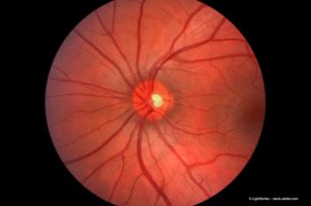
The questions surrounding idiopathic macular telangiectasia type 2, a disease that occurs more often than thought, is more severe than thought, and has confirmed that systemic associations, are slowly being answered as a result of the 5-year-old ongoing MacTel Project. However, information about disease pathogenesis remains elusive.
Mark C. Gillies, MB BS, PhD, FRANZCO, speaking for the MacTel Project Study Group, reported the status of the study during retina subspecialty day at the annual meeting of the American Academy of Ophthalmology.
Based on the population of the Beaver Dam Eye Study, the incidence of macular telangiectasia, characterized by loss of central luteal pigment and Müller cell markers in the central retina, was 0.1% of patients over 45 years, but this may have been an underestimation considering that only colour photographs were examined.
"This condition may be as common as or more common than retinitis pigmentosa," Dr Gillies said.
The severity of macular telangiectasia is also worse than previously suspected, with loss of cones and dense central scotomas resulting in paracentral scotomas that cause significantly greater dysfunction than is found in patients with age-related macular degeneration (AMD) with similar visual acuity. In particular, the general vision, distance vision and near vision were reported as worse by patients with macular telangiectasia compared with those with AMD, and these patients have a higher prevalence of diabetes, hypertension, obesity and coronary artery disease.
Familial transmission of the disease is recognized, but the gene has not yet been identified. Of the 500 enrolled participants in the study, 24 families have been identified with another family member affected, mostly sibling pairs. A 3.5-megabase region on chromosome 1 has been identified with a LOD score of 3.45, indicating the likelihood of a genetic defect.
Clinical scientists working on the project have found that autofluorescence is the most sensitive and specific marker for macular telangiectasia type 2.
No treatment has been established. Laser treatment is not helpful. Anti-vascular endothelial growth factor (VEGF) drugs for subretinal neovascularization can be used for subretinal neovascularization and these patients can do well, according to Dr Gillies.
Some research has been done in Germany to test the efficacy of anti-VEGF drugs administered before the development of subretinal neovascularization. The effect on angiographic hyperfluorescence in patients without subretinal neovascularization was observed after monthly injections of ranibizumab (Lucentis, Genentech).
"The treatment worked, and after 12 injections there was little fluorescence visible, but it recurred," he said. "These patients would likely need to be treated indefinitely to maintain the effect. The concern was the development in one treated eye of a very dense scotoma in the area where macular telangiectasia type 2 almost always starts, which is about 600 µm temporal to the center of the fovea and just beneath the horizontal midline. This scotoma might have been due to a neural toxic effect of VEGF inhibition. The recommendation was to stop treatment."
Causes of the disease are speculative. Dr Gillies suggested that it may result from Müller cell injury.
"The retinal damage may then proceed down a vascular pathway, with angiographic leakage and eventual subretinal neovascularization," he said. "The retinal damage could also proceed down a neuronal pathway independently or concurrently, with increased reflectivity of the outer nuclear layer leading to inner and outer retinal cavitation."
Dr Gillies and colleagues created a murine Müller cell knockout model. The mice developed macular telangiectasia 12 weeks after selective knockout of Müller cells.
The apoptosis in the outer nuclear layer was inhibited by injection of recombinant ciliary neurotrophic factor (CNTF). Exploratory trials of candidate therapies are considered for 2011.
Study of subclinical disease, especially with adaptive optics, will probably help determine the underlying disease mechanism. The project will conduct additional research to identify related gene defects, develop other animal models, use adaptive optics to study photoreceptor changes in the earliest clinical phenotype of the disease, as well as identify potential treatments.
Dr Mark C. Gillies, MB BS, PhD, FRANZCO is scientific manager of the MacTel Project and professor of ophthalmology at the Save Sight University of Sydney, Australia. He can be reached by E-mail:
Dr Gillies has no financial interest in this research. The research was supported by the Lowry Medical Research Institute.
Newsletter
Get the essential updates shaping the future of pharma manufacturing and compliance—subscribe today to Pharmaceutical Technology and never miss a breakthrough.


