Why the future of ophthalmology is in corneal grafts
Restoration and regeneration represent a new paradigm
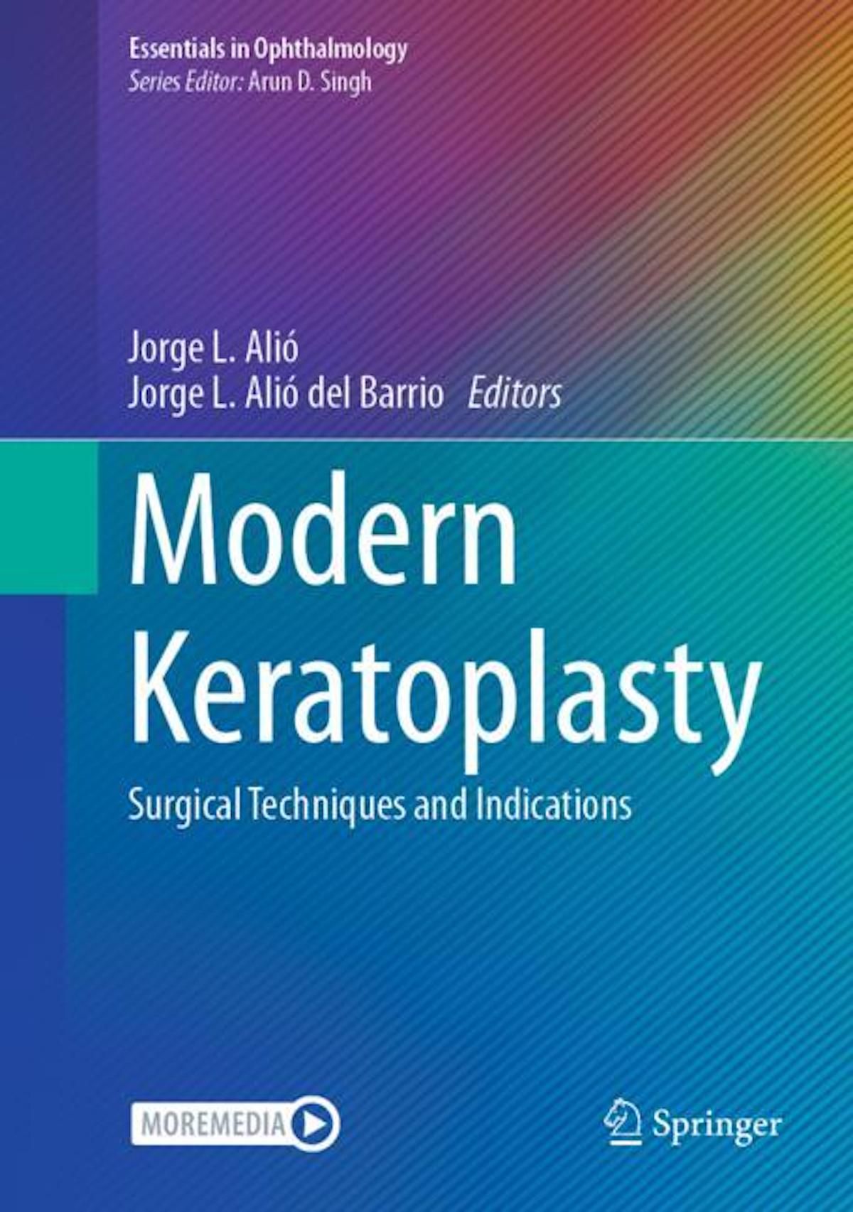
In a new book, Jorge L. Alió, MD, PhD, FEBOphth, and Jorge L. Alió del Barrio, MD, PhD, FEBOS-CR, FWCRS, explore the rapidly changing field of keratoplasty. In this excerpt from the book, our authors glance toward the future of corneal graft surgery.
"Modern Keratoplasty: Surgical Techniques and Indications" is available as part of the Essentials in Ophthalmology series from Springer Nature.
Image used with permission from Springer Nature.

Excerpt: Corneal Graft Surgery Provides a Glance to the Future
Throughout the book "Modern Keratoplasty," we witness the major evolution that corneal graft surgery has experienced over the last 2 decades. A better understanding of the corneal anatomy and physiology, technical improvements in the management of corneal bank tissue, improvements in surgical instrumentations (such as the availability of femtosecond laser), new surgical techniques that have emerged and have finally been consolidated as better options to the classical penetrating keratoplasty with better results, medical education and, above all, the skills and the talent of corneal surgeons have made corneal surgery enter a final stage of development since its early beginnings with the description of penetrating keratoplasty (PKP) by Zinn in 1906 and popularised by Castroviejo in 1936. Over all these years, the evolution has been constant and always for the benefit of better techniques, better results and better solutions to corneal blindness.
However, even though the results have been widely implemented, we see in the early chapters of the book how corneal graft still offers a challenge. Anatomical success does not always happen and anatomic failures are relatively frequent, with reported levels of survival from 52% to 98.8% for PKP at 10 years, from 77% to 99.3% for deep anterior lamellar keratoplasty at 5 years, from 56% to 94.1 % for Descemet stripping endothelial keratoplasty at 5 years and from 90% to 97.4% for Descemet membrane endothelial keratoplasty at 5 years.1 The main pitfalls are immune graft rejection, comorbidities and relapse of the previous disease.
In addition, functional failures, not frequently estimated as real ones, happen in a considerable number of patients, especially in PKP (for example, 10% recurrence of the ectatic disease at the host remnant peripheral cornea after 20 years), leading to a lack of adequate gain of visual acuity.2 Targeting the control and resolution of these problems, especially immune graft rejection, is mandatory and one of the real challenges that the modern corneal graft surgeons face. However, this is not going to be enough, as functional failures still influence the outcomes, and they are not always within the surgeon’s control. So, it seems mandatory to move to a totally different model, a paradigm shift. The philosopher Thomas Kuhn defined a paradigm shift as needing to happen first in the mind of the decision-makers in that particular topic.3 This is exactly what has to happen now in corneal surgery; we need a paradigm shift.
The new paradigm will be, instead of tissue replacement, tissue restoration by regeneration. Corneal regeneration is also targeted in the book, and it is still in its early stages. The possibility of restoring the ocular surface has been a highlight. The challenges that are involved are clear and there is still time ahead to achieve this on a consistent and repeatable basis. Translational research in this area is evolving, and in the coming years, it will probably take an important role in our practice once costs associated with it fade, allowing their availability to be more general. Corneal stroma regeneration, according to data from our recent pioneering clinical studies, seems to be feasible and even more easily applicable with the use of xenogenic tissue (instead of human corneal tissue, that is so scarce and expensive).4-7 Corneal endothelial substitution seems to be feasible even though the lack of biological productivity of the corneal endothelium seems to be a limitation during the culture of these cells.
Future clinical research, both basic and translational, will likely increase the efficiency and involved costs of such procedures in order to succeed in real clinical practice. We can clearly foresee that it, in the future eye cellular eye banks, containing the best donor lineages for each cell type, will centralise the production and delivery of the different stem cell regenerative products among all clinical centers, making these new procedures cost-effectiveand available for all ophthalmology clinics, without the need for investing in expensive facilities. These cells and derived tissues may no longer depend on ocular human sources at some point and start coming from human bioengineering extraocular sources or even xenogenic tissues.
Stem cells that contain proven immunomodulatory properties, will unlikely be provided from the same patient, affected by the same genetic imbalance that made the corneal disease to happen initially (except for those abnormalities caused by external aggressions), but rather from considered genetically 'optimal' donors in which immortal stem cells constantly proliferating will provide an easy and cheap way to restore the biology of the cornea.
Corneal graft surgery, as it is practiced today, it will never disappear and indeed will continue evolving, but it may be finally largely substituted by corneal regeneration procedures. We hope that the reader of our book will contribute to the medical education of those surgeons interested in corneal graft surgery and stimulate them to search for new innovative and better solutions for corneal blindness for the benefit of our patients.
References
1. Alió JL, Montesel A, El Sayyad F, Barraquer RI, Arnalich-Montiel F, Alio Del Barrio JL. Corneal graft failure: an update. Br J Ophthalmol. 2021 ;105(8): 1049-58. https://doi.org/10.1136/ bjophthalmol-2020-316705.
2. de Toledo JA, de la Paz MF, Barraquer RI, Barraquer J. Long-term progression of astigmatism after pen-etrating keratoplasty for keratoconus: evidence of late recurrence. Cornea. 2003;22(4):317-23.
3. Kuhn T. The Structure of Scientific Revolutions. 2nd ed. Chicago, IL: University of Chicago Press; 1970. ISBN 978-0-226-45804-5.
4. Alió Del Barrio JL, Arnalich-Montiel F, De Miguel MP, El Zarif M, Alió JL. Corneal stroma regeneration: preclinical studies. Exp Eye Res. 2021;202:108314. https://doi.org/10.1016/j .exer.2020.108314.
5. Zarif ME, Alió del Barrio JL, Arnalich-Montiel F, De Miguel MP, Makdissy N, Alió JL. Corneal stroma regeneration: new approach for the treat-ment of cornea disease. Asia Pac J Ophthalmol (Phila). 2020;9(6):571-9. https://doi.org/10.1097/ APO.0000000000000337.
6. El Zarif M, Alió JL, Alió Del Barrio JL,et al. Corneal stromal regeneration therapy for advanced keratoconus: long-term outcomes at 3 years. Cornea. 2021;40(6):741-54. https://doi.org/10.1097/ ICO.0000000000002646.
7. Alió del Barrio JL, De la Mata A, De Miguel MP, et al. Corneal regeneration using adipose-derived mesenchymal stem cells. Cell. 2022; 11 (16):2549. https://doi.org/10.3390/ cellsl 1162549.
8. Alió JL, Alió del Barrio JL, eds. Modern Keratoplasty: Surgical Techniques and Indications. Springer Nature; 2023. Essentials in Ophthalmology; vol 18. Accessed November 16, 2023. https://doi.org/10.1007/978-3-031-32408-6
Jorge L. Alió MD, PhD, FEBOphth | E: jlalio@vissum.com
Prof Dr Alió is Professor and Chairman of Ophthalmology, Miguel Hernández University of Elche, Alicante, Spain. He is the founder of Vissum Miranza, Alicante, Spain, and the Jorge Alió Foundation for the Prevention of Blindness.
Jorge L. Alió del Barrio, MD, PhD, FEBOS-CR, FWCRS
Jorge L. Alió del Barrio received his PhD from the University of Alcalá de Henares, Madrid, Spain. He is a cornea, cataract, ocular surface and refractive surgeon at Vissum, Miranza Group, Alicante, Spain. He is associate professor in the Department of Ophthalmology in the Faculty of Medicine of the Universidad Miguel Hernández in Alicante.

Newsletter
Join ophthalmologists across Europe—sign up for exclusive updates and innovations in surgical techniques and clinical care.