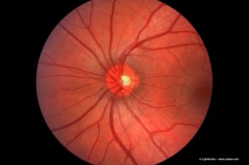
Triamcinolone acetonide: recommended for DME treatment?
Use of intravitreal triamcinolone acetonide (IVTA) has increased significantly over the past four years as a consequence of successful reporting of the agent's efficacy in the treatment of cystoid macular oedema resulting from uveitis, birdshot retinochoroidopathy, central retinal vein occlusion and diabetic macular oedema.1-6
Many clinical and experimental studies have convinced us of the lack of toxicity associated with intravitreal cortisone injection2,4,7,8 and testing in human eyes has allowed us to establish concentration and duration of effect.9-11 Pharmacokinetic studies on rabbit and human eyes receiving an intravitreal injection of triamcinolone have confirmed that the drug is delivered rapidly to its site of action with maximal bioavailability whilst remnants of the intravitreally injected suspension are visible for 21 to 41 days.12,13 We also know that the elimination half-life in human non-vitrectomized eyes after a single injection of triamcinolone is 18.6 days, with a measurable concentration in the vitreous humour lasting three months.14
Findings from a six-month study
Within the study group, one group had recurrent or persistent macular oedema despite previous macular laser treatment, whilst patients in the second group had never received laser treatment.
We prospectively considered 23 eyes of 22 patients with CSDME according to ETDRS criteria. Twelve eyes were refractory to macular laser treatment (group 1), and 11 eyes received IVTA as primary therapy (group 2). CSDME had persisted for more than three months but less than one year in all patients.
Eyes with previous vitrectomy, uveitis, laser treatment for choroidal neovascularization, history of glaucoma or macular thickness less than 300 microns by optical coherence tomography (OCT) were excluded from the study.
After a complete ocular examination, including best-corrected Snellen VA and macular thickness measured by OCT 3 (Humphrey Instruments, San Leandro, California, USA), a dose (4 mg, 0.1 mL) of triamcinolone acetonide (TA) (Kenacort 40 mg, Bristol-Myers Squibb Co., New Jersey, USA) was injected slowly through the temporal pars plana, 3.5 mm posterior to the limbus. All 23 eyes completed six months of follow-up.
Best-corrected VA and OCT examinations were performed prior to IVTA administration, 48 hours postinjection (first timepoint), every seven days post-IVTA for one month (second, third, fourth and fifth timepoints), at three months (sixth timepoint) and at six months (seventh timepoint) following the single injection.
Newsletter
Get the essential updates shaping the future of pharma manufacturing and compliance—subscribe today to Pharmaceutical Technology and never miss a breakthrough.


