The “fourth dimension” of home monitoring
A longitudinal, AI-based approach to fluid quantification
As home monitoring technology improves, so too does the range of applications. The data produced by at-home retinal monitoring, when paired with artificial intelligence (AI) analysis, may provide a scaffold for new understandings of retinal disease. This was the topic explored by Anat Loewenstein, MD, professor and director of the Department of Ophthalmology at Tel Aviv Sourasky Medical Center, in her presentation at the 21st annual Angiogenesis, Exudation, and Degeneration 2024 Conference. Her lecture, “Longitudinal AI-Based Fluid Quantification and Its Implication for Remote Patient Monitoring,” focused on home monitoring as it applies to patients with neovascular age-related macular degeneration (nAMD).
In her presentation, Prof Loewenstein described the advent of optical coherence tomography (OCT) as allowing ophthalmologists to visualise the retina in three dimensions. But home OCT, she said, introduces the “fourth dimension,” temporality.1 To better understand disease dynamics, practitioners need to assess biomarkers over time, and in real-world scenarios, not exclusively during office visits. By applying AI-based fluid quantification, Prof Loewenstein said, ophthalmologists can understand how temporal changes may reflect new disease dynamics and need for a modified treatment response.


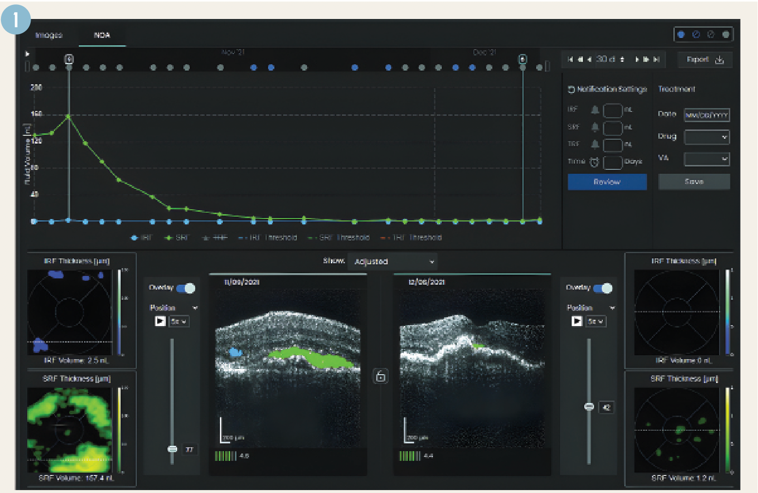
Figure 1. Notal OCT Analyzer Showing Fluid Volume Trajectory, B-Scans and Fluid Thickness Maps.
Prof Loewenstein shared a retrospective analysis of data collected via at-home OCT scans of 180 patients with 296 eyes. All patients had at least one eye diagnosed with nAMD. The mean age was 77.1 years; 57% of patients were female. The mean best corrected visual acuity was 20/37.
The patients received a Notal Vision home OCT device, which they self-installed and self-calibrated, with the option to receive help over the phone. During the 5-week study period, each patient performed, on average, 6.2 scans per week.
The patient cohort generated a total of 9,438 self-images in the period allotted. Daily scans were automatically synced to cloud storage and assessed by the Notal OCT Analyzer (NOA), which resulted in a physician report (Figure 1), viewable on the web. Data in the report included calculated fluid volume by type, fluid volume trajectory by fluid type, fluid thickness maps and raw and segmented B-scans.


Table 1. Categories & Prevalence
All categories show a meaningful prevalence in a 5-week study
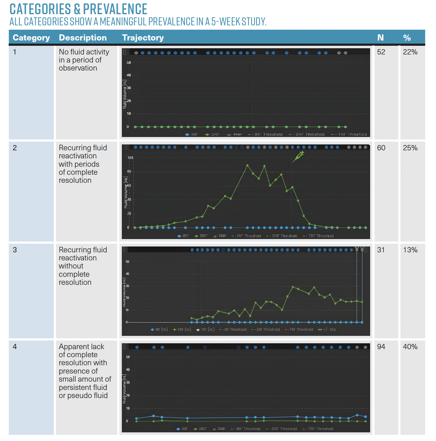
After images were captured, all nAMD eyes were categorised based on the patterns of the fluid trajectories (Table 1). Prof Loewenstein noted that this is the first attempt to categorise nAMD using temporal patterns.
Investigators developed four “meaningful categories” based on disease activity: eyes which showed no fluid activity during the period of observation; eyes which saw recurrence of fluid activation, with complete resolution after treatment; eyes which demonstrated periodic recurrence of fluid activation, but did not achieve full fluid resolution after treatment; and eyes with small amounts of stable persistent fluid, or pseudo fluid structures (such as degenerative cysts and outer retinal tubulations) and no complete resolution.1
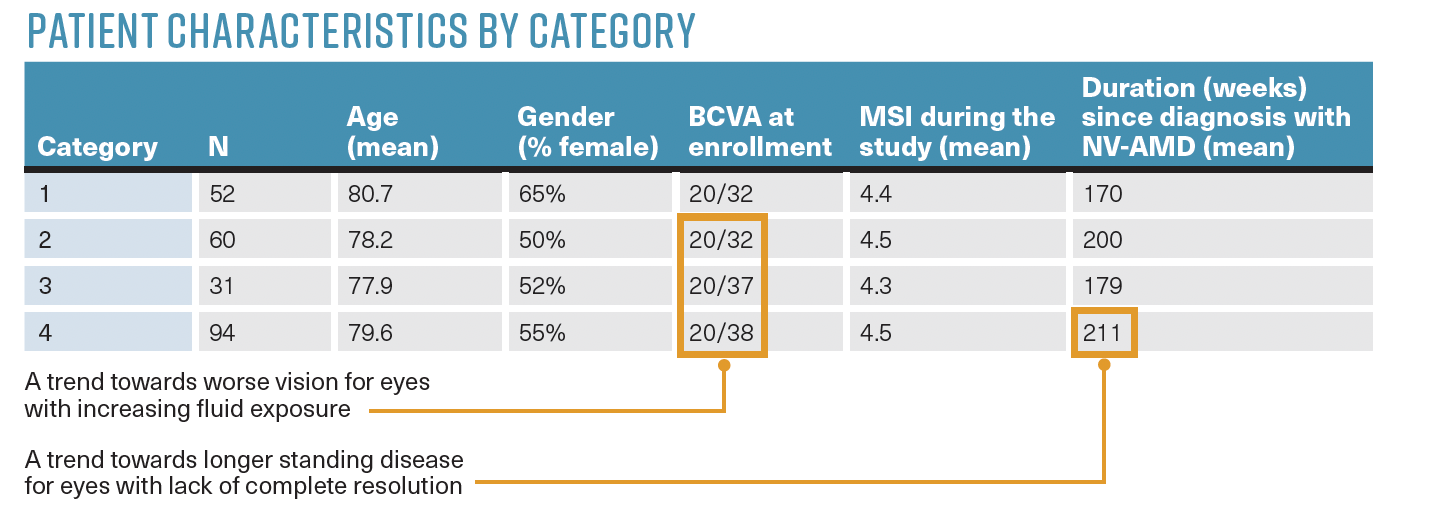
Upon reviewing the patient characteristics by category, Prof Loewenstein noted key points and trends. She noted that patients experienced slight worsening of vision as fluid exposure increased, or as patients developed pseudo fluid-like structures.
Eyes with longer-standing diagnoses of nAMD were more likely to exhibit persistent fluid or pseudo fluid, and to see a lack of complete resolution.
Along with providing an understanding of disease progression, the four patient categories also helped investigators develop patient management considerations (Table 2). Prof Loewenstein recommended that patients in the first category, with no fluid activity, should be observed and brought in for regular evaluation and, if disease activity presented, evaluated and treated accordingly.


TABLE 2. Patient Characteristics by Category.
Patients who fell into the second category, showing recurring fluid reactivation with periods of complete resolution, should be treated promptly if fluid reactivates. In certain cases, practitioners should consider preemptive therapies to prevent recurrence.
Patients in the third category, who experience recurrent fluid reactivation and no complete resolutions, may be candidates for novel therapies. Retina specialists should consider switching therapies for those patients under their care, to obtain and, ideally, maintain better fluid control.
Finally, patients in category four, who experience a lack of complete resolution and experience minimal persistent fluid or pseudo fluid, should remain under close observation. There may be no need for treatment, as long as fluid does not see significant increase.
Case study
To demonstrate the application of this categorisation, Prof Loewenstein presented a case study from a 73-year-old man monitored using home OCT. The patient’s left eye exhibited “category 3” characteristics; that is, the eye did not achieve full fluid resolution after multiple treatments.
The patient began home OCT in January 2023 (Figure 2). During the monitoring period, the patient experienced reactivation of intraretinal fluid with foveal involvement.

")
Figure 2. Case Showing Fluid Trajectory of Category 3 Patient. (Images courtesy of Anat Loewenstein, MD)
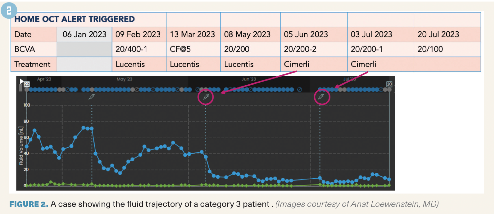
The patient was treated with
ranibizumab following fluid detection. Home monitoring captured a reduction in fluid, but not complete resolution. Prof Loewenstein noted that while the patient had a visual acuity (VA) of 20/400, he was still able to consistently self-image using the home OCT device.
The patient experienced another round of activation and was treated with the same drug, again with minimal resolution. Following a subsequent reactivation, the patient was treated with a different agent, ranibizumab-eqrn (Cimerli), a biosimilar to ranibizumab.1 Following this switch, the patient presented a faster and greater resolution in fluid, as well as VA improvement. Home OCT captured details on the change in fluid dynamics and treatment response.
Key takeaways
Prof Loewenstein said that home OCT is especially vital as patients face longer intervals between evaluations and between treatments. Retina specialists may not see patients for as many as 4 months between injections. By implementing home monitoring, practitioners can achieve a more comprehensive view of disease progression and treatment response.
Just as nAMD subtypes are classified with multimodal imaging, home OCT makes further classification possible, Prof Loewenstein added. Among the study cohort’s high density of data output, distinct trajectory patterns presented. Analysing that data can provide structure for highly personalised treatment plans and robust management guidelines for nAMD.
Reference
1. Loewenstein A. Longitudinal AI-based fluid quantification and its implication for remote patient monitoring: setting the stage for understanding biomarkers in the 4th dimension of OCT imaging. Lecture presented at: Angio-genesis, Exudation, and Degeneration 2024; February 2-3, 2024; Virtual.
Anat Loewenstein, MD | E: anatl@tlvmc.gov.il
Loewenstein is director of the Department of Ophthalmology, Tel Aviv Medical Center, Israel. She is a consultant to Notal Vision.


Newsletter
Join ophthalmologists across Europe—sign up for exclusive updates and innovations in surgical techniques and clinical care.


 and the International SPECTRALIS Symposium – And Beyond (ISS) in Heidelberg, Germany.")


")


 and Roland Rocholz, PhD, technology and innovation manager. Image courtesy of Heidelberg Engineering")