|Articles|May 1, 2011
Technique enables surgical success in IFIS
Combining 1.8 mm C-MICS and phacoemulsification system enables safe and effective cataract surgery in eyes with intraoperative floppy iris syndrome.
Advertisement
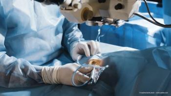
The combination of 1.8 mm coaxial micro-incision cataract surgery (C-MICS), proper wound construction, and attention to other elements of surgical technique aiming to maintain chamber stability allows safe and effective cataract surgery in eyes with intraoperative floppy iris syndrome (IFIS) without the need for pupil expansion, according to Dr John D. Hunkeler.
Key components of his technique include the creation and maintenance of snug trapezoidal incisions, use of a smaller capsulorhexis than usual, and a divideandconquer approach.
"The divide-and-conquer technique keeps the phaco tip behind the iris during nuclear disassembly to reduce the likelihood of iris trauma," he said. "Nuclear fragments are elevated into the anterior chamber by the phaco tip in aspiration mode for subsequent ultrasound removal, well away from the iris. 1.8 mm C-MICS with the [phaco] platform delivers reliable control of both phaco power and vacuum to maintain excellent chamber stability."
Less need for expansion devices
"Using this procedure I have been able to perform cataract surgery safely and successfully in eyes with IFIS, with no iris or capsule complications and without the need for pupil expansion devices in most cases," Dr Hunkeler added.
He reported that in 2008, when performing 2.8 mm coaxial phaco, he placed a pupil expansion device (Malyugin ring, MicroSurgical Technology) in 33% of IFIS cataract surgery cases. In 2009, his usage of the device in IFIS cases decreased to 9%, but those cases were mostly performed during the first half of the year.
"Since August 2009, I have only used the ring in a single IFIS cataract surgery case," he said.
Trapezoidal incision, paracentesis
Dr Hunkeler said he creates a clear corneal trapezoidal incision and a trapezoidal paracentesis, employing a paracentesis blade first to create the primary incision and then a triangular blade to create the trapezoidal component.
He said he also instills Shugarcaine and Amvisc Plus to help maintain maximum pupillary dilation throughout the procedure.
Using the divide-and-conquer technique, because it reduces the possibility of iris prolapse, he performs as much sculpting and cracking as possible in the posterior chamber before elevating the lens material to just above the pupillary plane, where it is removed with C-MICS phaco and irrigation/aspiration.
Once lens removal is completed, viscoelastic is instilled into the posterior chamber, and Dr Hunkeler implants a spherical, aberration-free microincision lens (Akreos MICS IOL, Bausch + Lomb) through the 1.8 mm clear corneal incision.
Special ContributorDr John D. Hunkeler is medical director at the Hunkeler Eye Institute, Kansas City and is the former chairman of the department of ophthalmology at the University of Kansas School of Medicine, Kansas City, Kansas, USA. He can be contacted via Email:
Dr Hunkeler is a consultant to Abbott Medical Optics and Bausch + Lomb.
Advertisement
Related Content
Advertisement


Latest CME


Advertisement
Advertisement
Trending on Ophthalmology Times Europe
1
Demodex blepharitis: Closing the diagnostic gap to improve ocular surface health
2
Mulling over options for mitigating severe vision loss from diabetes
3
Trehalose-hyaluronate combination aids tear film after eyelid surgery
4
ZEISS reports early implantations of AT LUCIA Toric 721P IOL in Europe
5