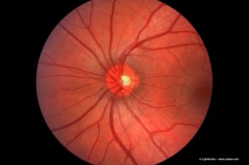
Submacular surgery: the viable alternative
Dr Claus Eckardt discusses the alternative to anti-VEGF drugs, to be employed when other measures have failed.
Key Points
Today, in the anti-VEGF drug era, macular surgery is indicated only in cases in which no improvement is expected with administration of ranibizumab (Lucentis; Novartis) or bevacizumab (Avastin; Genentech), such as in eyes with tears in the retinal pigment epithelium (RPE), large subretinal haemorrhages, or in eyes in which the VA has decreased despite the use of anti-VEGF therapy.
"Surgical therapy for age-related macular degeneration (AMD) provides some improvements in visual acuity (VA) for patients whose conditions do not respond to anti-vascular endothelial growth factor (VEGF) drugs," said Claus Eckardt, MD.
The three approaches
Macular translocation
"In another case, a 78-year-old patient presented with a haemorrhage that had occurred only one hour previously. We performed macular translocation surgery one day later. Sixteen months later, VA had improved from 0.1 to 0.8 in the better eye. The patient was able to return to medical practice six weeks after the silicone oil was removed.
"I am not aware of any technique other than translocation surgery that would be more successful at repairing rips in the RPE," he said.
A report of eight cases published by Yusuke Oshima, MD, PhD, and colleagues from the Osaka University Graduate School of Medicine and Faculty of Medicine in Japan1 described how the authors drained the liquid blood from eyes 24 hours after the injection of tPA. They drained the blood through two small peripheral retinotomies by injecting perfluorocarbon liquid onto the retina. At a follow-up point of two years, VA had improved significantly in all eyes but one.
Peripheral retinotomy
"My group performs a large peripheral retinotomy (~250°)," Dr Eckardt recounted. "They then reflect the retina to remove the blood and fibrovascular proliferation. In the presence of a small RPE defect, the retinotomy is enlarged to 360°, and macular rotation is performed.
"The procedure is easy to perform if the haemorrhage is not fluid; if the haemorrhage is fluid, then performing the retinotomy can be difficult because the blood may obscure visualization."
In a patient who presented eight months after reading vision was lost due to submacular choroidal neovascularization, two months after a haemorrhage that further decreased vision, and after six intravitreal bevacizumab injections, Dr Eckardt removed the subretinal blood via a 250° peripheral retinotomy.
"About 18 months after the procedure, the vision is 0.2, and the foveal fixation has been regained. In addition, the patient was able to return to work one week after the procedure," Dr Eckardt said.
"Our results with intravitreal tPA and gas injection have not been good," Dr Eckardt acknowledged, "but still we attempt the procedure."
Newsletter
Get the essential updates shaping the future of pharma manufacturing and compliance—subscribe today to Pharmaceutical Technology and never miss a breakthrough.


