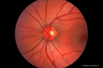
Spontaneous closure of FTMH after vitrectomy for VMT
In this article, the authors summarize a recently published case study of a macular hole formation and its spontaneous closure after vitrectomy.
In our clinic we experienced a case of a macular hole formation and its spontaneous closure after vitrectomy for VMT, which we have recently published.1 In this article, we will present a brief summary of this case report and our findings.
Case presentation
A week after vitrectomy we found, using SD-OCT examination (Spectralis, Heidelberg Engineering, Heidelberg, Germany), that the VMT had resolved but a full-thickness macular hole (FTMH) had formed. A month later the FTMH had spontaneously closed, and after another 5 months, SD-OCT showed a normal foveal contour.
First description
The current case is, to the best of our knowledge, the first description of macular hole formation and spontaneous closure after vitrectomy for VMT that is clearly documented step-by-step with SD-OCT.
The formation of FTMH may have been a result of the creation of anterio-posterior traction on the fovea by the posterior hyaloid membrane, in addition to, damage caused to the retinal layer in the macular area during detachment.
In this case, the traction was pulled up to the retina, so that the edges of the hole were elevated high, but it was stabilized by the ILM and the posterior hyaloid membrane during surgically induced detachment. The complex of the posterior hyaloid membrane and ILM was probably removed in the macular area and FTMH developed.
Conclusion
FTMH may develop in its natural course and after vitrectomy for VMT.2,3 However, in these referenced cases there was no spontaneous closure of the FTHM, they all required further surgery to close the macular hole.
The release of the mechanical traction may be the main reason for the eventual closure of the macular hole. However, ILM peeling induces glial cell proliferation across the hole and this mechanism may also help the spontaneous closure of macular hole.
References
1. D. Odrobina et al., BMC Ophthalmology, 2014;14:17.
2. N. Yamada et al., Am. J. Ophthalmol., 2005;139(1):112–117.
3. D. Odrobina et al., Retina, 2011;31(2):324–331.
Newsletter
Get the essential updates shaping the future of pharma manufacturing and compliance—subscribe today to Pharmaceutical Technology and never miss a breakthrough.


