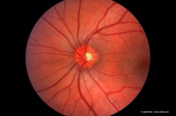
Simplified severity scale may be predictive of vision loss in patients with AMD
The Simplified Age-Related Eye Disease Study (AREDS) Age-Related Macular Degeneration (AMD) Severity Scale has been shown to be predictive of vision loss.
Key Points
"Fundus lesions can be used as outcomes in prevention studies for the development of advanced AMD and may alleviate the need for fluorescein angiography in large studies," said Dr. Chew. She is deputy director of the Division of Epidemiology and Clinical Research, National Eye Institute, National Institutes of Health, Bethesda, MD.
The well-known AREDS, which began in 1992, evaluated the clinical course and prognosis of AMD and lens opacities of 4,757 men and women who were aged 55 to 80 years at enrollment in a randomized, controlled clinical trial of antioxidant vitamins and minerals. The clinical trial ended in 2001, and 3,687 participants were followed until 2005.
The purpose of the presentation was two-fold: to assess the 10-year risk of visual acuity (VA) loss in participants who developed fundus lesions associated with advanced AMD and to assess the risk in each of the stages of the severity scale, Dr. Chew said.
The fundus lesions associated with neovascular AMD that were evaluated included serous detachments of the sensory retina, non-drusenoid retinal pigment epithelial (RPE) detachments, subretinal or sub-RPE hemorrhages, and subretinal fibrosis. Other lesion of advanced AMD evaluated was geographic atrophy that involved the center of the macula.
Various groups were identified within the study population:
"A total of 685 eyes had any neovascular AMD event," said Dr. Chew. "The first time neovascularization was observed, it could be one or more of the fundus lesions present in an eye." In those eyes, the baseline VA was 20/32. The VA level decreased to about 20/160 at the 5-year time point and to about 20/300 at the 10-year time point.
Further analyses then evaluated each fundus lesion separately. In the group of 519 patients who had a serous subretinal or hemorrhagic retinal detachment, the median VA at baseline of 20/32 declined to about 20/200 at the 5-year examination and about 20/300 at the 10-year time point, Dr. Chew said.
In 302 patients with subretinal fibrosis, a more rapid decline in VA was seen over time, with the median baseline VA dropping from 20/40 to 20/320 at the 5-year time point and again at the 10-year time points.
The development of central geographic atrophy (428 eyes) resulted in the VA decline from a median baseline value of 20/40 to 20/160 at the 5-and 10-year time points.
Newsletter
Get the essential updates shaping the future of pharma manufacturing and compliance—subscribe today to Pharmaceutical Technology and never miss a breakthrough.


