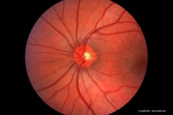
The role of fundus autofluorescence is expanding
Technology is an essential tool for diagnostic, prognostic approaches to AMD management
FAF provides a topographic map of accumulated lipofuscin within the retinal pigment epithelium (RPE) and represents a non-invasive means of identifying pathologic changes that cannot be visualized with normal fundus photography, fluorescein angiography, or optical coherence tomography (OCT).
Lipofuscin is an indicator of photoreceptor turnover and its primary fluorescent component is A2E, a potentially toxic vitamin A metabolite implicated in the pathogenesis of AMD, said Dr Csaky, senior scientist, Retina Foundation of the Southwest, Dallas, USA.
A study by Schmitz-Valckenberg et al., published in 2008 comparing these two technologies found that visualization of FAF patterns was better using the cSLO unit, which is explained in part by the fact that the SLO system uses signal averaging, generating a mean image from multiple scans that amplifies the FAF signal. Since that study was conducted, an updated version of the fundus camera system, which incorporates new filters, has been released.
According to the manufacturer, the modification has improved the system's sensitivity, although that claim has yet to be verified in a clinical trial. The cSLO technology retains a clear advantage for better performance in eyes where there is lens opacity, Dr Csaky said.
FAF has been found to have a useful diagnostic application for helping to discriminate between AMD and certain retinal dystrophies that have a similar appearance on OCT and fluorescein angiography.
Dr Csaky also explained that the subretinal deposits in eyes with retinal dystrophies tend to have a high level of autofluorescence. Therefore, the FAF signal generated when those disorders are present is much more intense than that associated with AMD.
Ongoing research is trying to determine whether the various FAF patterns (speckled, defect, butterfly) that have been described in eyes with early AMD are predictive of the risk of vision loss from progression to geographic atrophy (GA) or exudative disease.
"So far we only know that patients with early dry AMD with drusen manifest with a variety of AF patterns," he said. "The prognostic implications await further study."
AF imaging may also have predictive utility in eyes with wet AMD whereas a marker of RPE viability, it may provide information on visual prognosis. In this regard, the results of one study indicate that maintenance of a normal FAF signal under the fovea suggests the capacity for vision to improve with anti-VEGF therapy.
Preventing progression of disease
FAF imaging is also being used as an outcome endpoint in research studies of novel investigational agents being tested for preventing the progression of early AMD.
"We know that disease progression occurs very slowly," Dr Csaky said. "And so it can take many years to determine the efficacy of a therapy if the endpoint is vision loss.
"However, it remains to be determined whether AF imaging, fundus photography, or fluorescein angiography provides a better marker for evaluating the area of GA and its progression," he added.
FAF may also be useful for following patients with GA because there is evidence that increased rim autofluorescence around an area of GA is a marker for GA progression. Confirmation of this feature as a prognostic factor will be important for patient counselling and in optimizing the selection of patients for inclusion in clinical trials.
"Interventional studies, including patients whose disease is likely to progress more quickly, will facilitate earlier identification of treatment efficacy," Dr Csaky concluded.
Newsletter
Get the essential updates shaping the future of pharma manufacturing and compliance—subscribe today to Pharmaceutical Technology and never miss a breakthrough.


