
|Articles|March 1, 2014
The refractive surgeon's secret to enhanced success
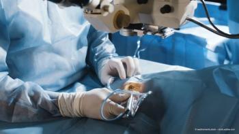
In this article Dr Kanellopoulos shares his opinions on AS-OCT in his practice, highlighting the clinical and diagnostic advantages he has experienced using the imaging device.
Advertisement
The beauty of recent developments in the AS-OCT that my colleagues and I have chosen for our practice (RTVue, Optovue Inc., Fremont, California, USA) is that it allows for an extremely simple examination that does not require a highly skilled examiner. It is user-friendly and, like other diagnostic devices we choose for our office, has point-and-shoot simplicity with high reproducibility. Optovue has recently launched a new, highspeed OCT, Avanti, and in my opinion, the widefield imaging and scan speed available on that device is unparalleled, and the large screen size allows a clinician to detect pathology better than a limited scan size.
The clinical science of a spectral domain OCT epithelial imaging comes into play when making surgical decisions not only postoperatively but also preoperatively. For example, it is commonly useful when a patient needs to be further evaluated for suspicious topography. Dry eye, blepharitis, contact lens use and even overmedication with drops in the office, may interfere with the intact epithelial distribution.
An epithelial map may be the 'missing' part of the puzzle when making decisions for refractive surgery. If a patient's anatomy is normal, then we can rely more on other imaging devices that provide the basics, such as the curvature and the power of the different parts of the cornea. If the epithelium is abnormal, then one has to take that into account, first, because an abnormal epithelium can interfere with Scheimpflug imaging, meaning Scheimpflug imaging may become significantly biased by such changes. Additionally, if the epithelium is abnormal, the better way to document it is with an epithelial map.
It has been to our surprise that many mishaps in refractive surgery and IOL lens calculation in cataract surgery were related to our inability, up until now, to have a reproducible, easily attainable epithelial map in patients.
Advertisement
Related Content
Advertisement


Latest CME


Advertisement
Advertisement
Trending on Ophthalmology Times Europe
1
Review highlights gaps in transition from paediatric to adult eye care
2
Mulling over options for mitigating severe vision loss from diabetes
3
Trehalose-hyaluronate combination aids tear film after eyelid surgery
4
Beyond anti-VEGF: The next wave in retinal therapy
5