Optical coherence tomography
Dr Sebasiten Wolf describes how SD-OCT has improved the visualization of intraretinal morphologic feaures both quantatively and qualatively.
The OCT technique is an interferometric imaging technique that generates cross-sectional images by mapping the reflections of low-coherence laser light from different tissue layers. Spectral or Fourier domain OCT refers to Fourier transformation of the optical spectrum of the low-coherence interferometer. With the spectral domain technique, imaging with an axial image resolution of about 5–10 µm is possible. The transversal resolution is limited by the optics of the eye, by the low numerical aperture of the optics of the illuminating beam, and the number of A-scans used to reconstruct a B-scan.
Currently, various SD-OCT instruments are commercially available. All systems provide high-quality OCT line scans as well as special scan patterns for imaging the optic nerve fibre layer around the optic disc and for producing 3-dimensional OCT images.
Most of the new SD-OCT systems image the outer retinal layers as three hyper reflective bands. The innermost of these hyper reflective bands has the lowest reflectivity. The bands may correspond to the external limiting membrane, the junction of the photoreceptor OS and IS and the RPE.
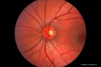
Various macular diseases have been studied and SD-OCT can aid in identifying, monitoring and quantitatively assessing various posterior segment conditions including diabetic macular oedema (DME), age-related macular degeneration (AMD), macular oedema (ME) as a result of retinal vein occlusion, disease of the vitreomacular interface such as epiretinal membranes, full-thickness macular holes, pseudoholes, schisis from myopia or optic pits, central serous chorioretinopathy, retinal detachment. At the same time it may be possible to explain why some patients respond to treatment while others do not. For example, SD-OCT is a valuable tool in determining the minimum maintenance dose of a certain drug in the treatment of DME, ME as a result of retinal vein occlusion (RVO) and wet AMD. It may demonstrate retinal changes that explain the recovery in some patients or non improvement in other patients without performing any invasive procedure such as retinal angiography.
Newsletter
Get the essential updates shaping the future of pharma manufacturing and compliance—subscribe today to Pharmaceutical Technology and never miss a breakthrough.


