|Articles|November 1, 2009
New 'Bimanual Rhexis' technique, for sub 1 mm incision, using the 'Capsulorhexis Chopsticks'
A new bimanual capsulorhexis technique, suing a specially developed instrument allowing a tri-dimensional, sub 1mm capsulorhexis with improved maintenance of the anterior chamber.
Advertisement
Key Points
The capsulorhexis device is a key driver for Micro Incision Cataract Surgery (MICS, sub 2 mm) and Micro-MICS (sub-1mm). MICS phaco techniques are gaining popularity but instrumentation for capsulorhexis is obviously a limiting factor.
There are two main capsulorhexis devices currently available:
Advantages
Drawbacks
2. Capsulorhexis Forceps
Advantages
They provide good and very precise control of the capsular tear. It's fast and simple to use, allowing a tri-dimensional control of the capsule. Surgeons can use any OVD either cohesive or dispersive.
Drawbacks
They are expensive and they are fragile. They need sterilization and for some of the models they cannot be use through sub 1 mm incisions.
So, what is the ideal capsulorhexis device and technique?
As far as the instrument is concerned it should be simple, inexpensive, solid, single-use and work through any incision size, even sub 1 mm incisions. With regard to the technique this should have only a short learning curve, be precise and reliable and allow a large capsulorhexis using any OVD. The control of the capsular tear should be tri-dimensional, without any OVD linkage. The anterior chamber (AC) space maintenance should be good even in case of shallow AC.
We are confident that this is achievable thanks to the 'Bimanual rhexis' technique, using a newly developed instrument, the 'Capsulorhexis Chopstick.'
Bimanual Rhexis technique
– Step 1
An initial sub 1 mm clear corneal incision is performed at 8 o'clock. Any OVD is injected inside the anterior chamber.
A second sub 1 mm clear corneal incision is then performed at 4 o'clock. Using the same knife, an anterior capsular tear is started at 6 to 8 o'clock.
– Step 2
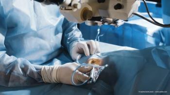
The two dedicated devices (Capsulorhexis Chopsticks) are introduced through both incisions.
Then the capsular tear is caught horizontally between the two chopsticks instead of the vertical plane of a standard forceps.
The capsulorhexis is performed in this horizontal plane using a shearing technique as opposed to the tearing technique used by some surgeons.
The capsular tear is grasped 3 to 4 times to complete the rhexis. The anterior chamber is very well maintained since there is no, or minimal OVD linkage through the two sub 1 mm incisions.
The Capsulorhexis Chopsticks
A specific device called The Capsulorhexis Chopsticks has been manufactured by Bausch & Lomb for this technique. They can be made of 100% stainless steel or a mix of steel and plastic for a single-use instrument. Both chopsticks are identical. The design has been specified to complement the technique. The distal part of the Chopsticks benefits from a surface treatment which enhances the adhesion with the capsule during the manoeuvres.
To summarise:
– Two perpendicular sub 1 mm clear corneal incisions
– Start the capsular tear between 6 and 8 o'clock at the final diameter (5,5 mm)
– Left Chopstick placed above the right, directing the capsulorhexis to 2 to 3 o'clock. The right Chopstick just follows.
– Bimanual transition at 2 o'clock
– Right Chopstick is placed above the left directing the capsulorhexis until the end. The left Chopstick just follows.
How widely has the technique been used?
Our experience is now based on more than 100 cases. The learning curve is short. The mean capsulorhexis diameter on the first 100 cases is 5,45 mm. The mean time is up to 28 seconds but this can be reduced dependent upon the surgeon's experience. All capsulorhexis procedures were completed successfully, without complications. The specific instrument design and surface treatment was able to achieve an excellent grasping of the anterior capsule.
Conclusion
We believe this new bimanual capsulorhexis technique using the Capsulorhexis Chopsticks is an effective and safe technique to perform a sub 1 mm tri-dimensional capsulorhexis. Any phaco or injection technique can follow. It represents a new philosophy of performing a capsulorhexis that enhances efficacy and safety thanks to a better maintenance of the anterior chamber.
Advertisement
Related Content
Advertisement


Latest CME


Advertisement
Advertisement