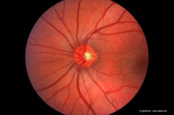
Navigated photocoagulation
This new technology will enable surgeons to perform treatments more accurately and comfortably in a short period of time.
Despite having a variety of imaging and treatment technologies available for retinal disease management, each technology has inherent properties that define its limitations. Integration of combinations of these technologies promises to be the best remedy for these individual shortcomings and may help achieve optimal outcomes.
Current slit lamp technology - while offering familiar functionality - is restricted in its limited field of view, which makes the task of viewing and evaluating pathologies more difficult. It is often challenging to locate the required landmarks in altered maculas using slit lamp viewing because of distortion or obscuration of key features.
A proprietary navigated photocoagulation system (Navilas, OD-OS) is an integrated system featuring real-time, widefield digital fundus imaging and retinal photocoagulation delivery. This advanced system has the capability to integrate imaging, planning, navigation, documentation and treatment delivery within a single device. The navigated photocoagulation system enables live wide angle viewing on a high-resolution monitor, which significantly surpasses the view obtained with slit lamp examination. This enables the surgeon to obtain a clean, crisp, non-inverted image of the whole macula at once, rather than having to rely on segmental fields-of-view. The clarity and resolution of the resulting image is comparable with that obtained with a scanning laser ophthalmoscope, but it has the additional advantage of colour, which improves the surgeon's evaluative capabilities.
In the past, static pre- and postoperative FA images would be placed side-by-side to compare the changes before and after treatment. Attempts have been made to overlay these FA images, but this has proven to be problematic owing to the fact that the images are rotated frequently and not easily aligned.
The solution provided by the navigated photocoagulation system is the use of high-speed tracking and computer registration of FA images onto the live fundus view of the patient. With the system's novel registration technology (navigation) - allowing the aiming beam to follow eye movement - there is increased confidence for the surgeon, who is subsequently able to target and deliver treatment more accurately in spite of any ocular movement intraoperatively.
Newsletter
Get the essential updates shaping the future of pharma manufacturing and compliance—subscribe today to Pharmaceutical Technology and never miss a breakthrough.


