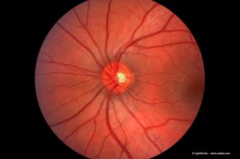
MCP-1 plays critical role in mediating photoreceptor apoptosis
Monocyte chemoattractant protein 1 (MCP-1) seems to be responsible for apoptosis of the photoreceptors in certain visual disorders.
Key Points
Joan W. Miller, MD, and colleagues demonstrated that MCP-1 has a critical role in mediating photoreceptor apoptosis after retinal detachment in an experimental murine model. Specifically, MCP-1 causes macrophages and microglia to accumulate and generate oxidative stress in the retina. She reported the findings of her group at the Retina Society meeting in Boston.
She is chairwoman, Department of Ophthalmology, Harvard Medical School, and chief of ophthalmology and Henry Willard Williams Professor of Ophthalmology, Massachusetts Eye & Ear Infirmary, Boston. The investigators reported their findings in the Proceedings of the National Academy of Sciences (2007;104:2425-2430).
"Because of this, new insights about the mechanisms that underlie photoreceptors' apoptosis in retinal detachment would be of clinical interest and could lead to new treatments," she said.
Suppression of caspases alone, which Dr. Miller and colleagues indicate is associated with death of photoreceptors as the result of retinal detachment, cannot stop apoptosis.
Dr. Miller explained that MCP-1 causes leukocytes to accumulate in diseases involving atherosclerosis, pulmonary infections, or angiogenesis, as well as some central nervous system disorders. In addition, MCP-1 is present in very high levels in vitreous samples from patients with retinal detachment compared with samples from patients with macular hole or idiopathic premacular fibrosis.
In the study under discussion, the authors used a murine model in which retinal detachments were induced in the right eyes of the mice. The animals received a subretinal injection of MCP-1 blocking antibody.
The authors found that the level of MCP-1 mRNA levels in the retina had increased 84-fold by 72 hours after the creation of the retinal detachments, and protein levels increased 10-fold compared with the baseline value.
"MCP-1 immunoreactivity was very weak in the normal controls, but spindle-shaped cells, which morphologically resembled Muller glia, were strongly MCP-1-immunoreactive in the outer plexiform layer after retinal detachment," they reported.
The results also showed that mice that are deficient in MCP-1 had significantly reduced infiltration of macrophages and microglia following the creation of retinal detachment and little photoreceptor apoptosis.
When the authors cultured primary adult retinal cultures to study whether increased MCP-1 is involved in photoreceptor death, MCP-1 was found not to affect the survival of the photoreceptors when the macrophages and microglia were removed from the cell cultures. They reported, however, that "without depletion of CD11b+ cells from the retinal cultures, the number of recoverin+ photoreceptors declined progressively with increasing MCP-1 concentrations, and MCP-1 concentrations as low as 0.1 mg/ml had significant cytotoxic effects. These data suggest that MCP-1's cytotoxicity is mediated through resident macrophage/microglia but not through direct interaction with the cultured photoreceptors."
In this experimental murine model of retinal detachment, Dr. Miller said, MCP-1 may be an important mediator of the macrophage and microglial responses in some retinal diseases. She pointed out that in a previous study of a retinal ischemia-reperfusion model conducted by Nakazawa and colleagues (Curr Eye Res 2003;26:55-63), MCP-1 was up-regulated in the inner retina only.
"In the current study, MCP-1 protein expression was detected in Muller cells, especially in the outer plexiform layer," Dr. Miller said. "The outer plexiform layer has a rich vascular capillary bed, which would facilitate extravasation of leukocytes."
Newsletter
Get the essential updates shaping the future of pharma manufacturing and compliance—subscribe today to Pharmaceutical Technology and never miss a breakthrough.


