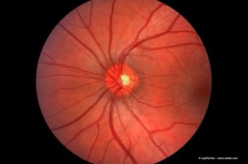
The evolution of the management of GRT

Article

During the advent of surgical approaches for retinal detachment repair, one of the most challenging cases a retinal surgeon may come across was a giant retinal tear. This article summarizes the history of GRT management and discusses current trends in surgical management.

The management of GRTs has evolved considerably over the decades. Early surgical techniques such as intraocular balloons (1970s), rotating tables and retinal incarceration with tacks, sutures and screws (1980s) have evolved into small gauge pars plana vitrectomy (PPV), wide field viewing systems and intraoperative perfluorocarbon liquid (PFCL) use. This article will briefly summarize the history of GRT management and discuss current trends in surgical management.

History of GRT management

Success rates in the management of GRTs are associated with the ability of the surgeon to manipulate the retinal flap back into its original configuration and maintain its position with intraoperative and postoperative tamponade. Earlier attempts were limited by an inability to effectively achieve and maintain the proper position of the retinal flap. A primary encircling scleral buckle could be successful in cases of minimally displaced GRTs with the placement of a low broad based scleral buckle. In the early 1980s, with the evolution of vitrectomy techniques, the surgeon could manipulate the GRT intraoperatively with rotating tables and intraocular gas tamponade.

Newsletter

Join ophthalmologists across Europe—sign up for exclusive updates and innovations in surgical techniques and clinical care.

Recent Videos
Christine Curcio, PhD, of the University of Alabama at Birmingham Heersink School of Medicine, shares histology update and revised nomenclature for OCT with Sheryl Stevenson of the Eye Care Network and Ophthalmology Times
SriniVas R. Sadda, MD, FARVO, shares key points from his retina presentation at the International SPECTRALIS Symposium
Robert Sergott, MD, describes fluorescence lifetime imaging ophthalmoscopy (FLIO) and the International SPECTRALIS Symposium – And Beyond (ISS) in Heidelberg, Germany.
Anat Loewenstein, MD, describes her presentation on remote imaging for age-related macular degeneration and geographic atrophy at the International SPECTRALIS Symposium, in conversation with Hattie Hayes of Ophthalmology Times Europe
Tyson Brunstetter, OD, PhD, a US Navy Aerospace Optometrist at the NASA Johnson Space Center in Houston, Texas, shares key takeaways from his keynote at the International SPECTRALIS Symposium (ISS)
Viha Vig, MBChB graduate student at the University of Auckland, New Zealand, discusses her poster presentation on the relationship between mitochondiral disease, Alzheimer disease, and other types of dementia.
Alfredo Sadun, MD, PhD, chief of Ophthalmology at the Doheny Eye Institute, University of California Los Angeles, shared exciting new research with the Eye Care Network during the Association for Research in Vision and Ophthalmology (ARVO) meeting on the subject of Leber hereditary optic neuropathy (LHON).
At this year's Association for Research in Vision and Ophthalmology (ARVO) meeting in Salt Lake City, Utah, Nitish Mehta, MD, shared highlights from his research documenting real-world results of aflibercept 8 mg for patients with diabetic macular oedema.
ARVO 2025: Anat Loewenstein, MD, shares data from herself and her colleagues on meeting needs of patients with diabetic retinopathy
A photo of Seville, Spain, with the Congress on Controversies in Ophthalmology logo superimposed on it. Image credit: ©francovolpato – stock.adobe.com; logo courtesy COPHy
© 2025 MJH Life Sciences

All rights reserved.