Evaluating subthreshold laser technique for CSCR patients
Ongoing study aims to clarify patient selection for endpoint management
Chronic CSCR patients who have less RPE scarring stand to benefit the most from EpM laser therapy. They have the best chance for a quick and complete resolution of subretinal fluid.
Among the more common retinopathies in the United States-along with
The exact etiology and pathogenesis is not well understood, but it has been reportedly associated with a range of factors such as corticosteroid exposure, phosphodiesterase inhibitor use, and obstructive sleep apnea.2-4 Interestingly CSCR has also been associated with “
An inciting event is thought to trigger an increased permeability of the choroidal vessels and
It has been reported that more than 80% of CSCR patients will have spontaneous resolution of symptoms within three months, nevertheless, the other 20% often require treatment.7-9 These individuals may have persistent serous macular detachment, vision loss, and subjective impairment.10,11
Although definitions of chronicity vary, a patient can be considered to have chronic CSCR if subretinal fluid has not resolved by three months. In practice, the classic patient with chronic CSCR presents with reduced visual acuity and contrast sensitivity, visual distortions, and a change in color vision.
RELATED:
CSCR presentation
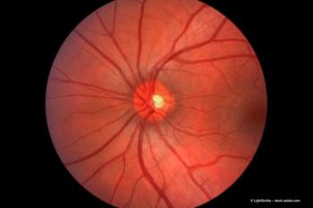
Clinically, the retina will have subretinal fluid and RPE irregularities that can also be seen with
In a very progressed form of the disease, the patient may have a complete loss of
To identify patients with the signs of secondary neovascularization, we take care to look for double layer signs on OCT and typical signs on fluorescein and ICG angiography.
A double layer of the RPE and Bruch’s membrane filled with hyper-reflective material is an indicator for
RELATED:
Treatment options
In the absence of a gold standard treatment, various strategies have been attempted in CSCR management. Specialists frequently apply laser photocoagulation to the leaking RPE, as directed by fluorescein angiography.
The development of subthreshold laser therapy has garnered growing interest as these techniques-including micropulse, selective retinal therapy, and
We find that PDT with reduced dose (half-dose) is the preferred approach for this method.16-19 Other drug treatments that have been used for CSCR include intravitreal anti-VEGF, mineralocorticoid receptor antagonists, adrenergic blockers, systemic carbonic anhydrase inhibitors, aspirin, Helicobacter pylori treatment, and methotrexate.20
RELATED:
Our investigation
My colleagues and I have initiated an investigation evaluating patients with chronic central serous chorioretinopathy, who are good candidates for EpM. Currently, we treat CSCR patients every three months if they present with subretinal fluid, a protocol that was also suggested by the technology’s inventors.
To see how suitable EpM is in the day-today practice of a clinical outpatient service, we have set up trial conditions to follow patients and gather more detail.
We will be looking closely at the anatomical outcomes on OCT as well as patients’ functional vision through visual acuity testing and microperimetry. We hope to include at least 50 patients. Currently, there are 30 are enrolled.
The follow-up period will be a year, and we would like to extend that out to longer intervals so we can examine recurrence after one year. We know that one of the difficulties in treating CSCR is that it is likely to recur with subretinal fluid-even after other therapies like half-fluence PDT.
We observe this on a regular basis. During our investigation, we want to determine which patients seem to improve the most and if there also are specific subgroups who will derive the most benefit from it.
Eventually, we will randomly assign patients to treatment and no treatment or treatment versus another type of treatment to show superiority. First, we want to evaluate the treatment’s effectiveness and improve our technique with the procedure. We want to ensure it does no harm. We had observed no adverse effects,
RELATED:
Conclusions
Through our observations, we believe that chronic CSCR patients who have less RPE scarring stand to benefit the most from
Patients with leakage points within a 3-mm radius from the fovea appear to do well with EpM’s macular laser pattern. Patients who have chronic CSCRs with fluid accumulation in multiple locations are more difficult to treat. An extended recruitment period and a longer follow-up will allow us to include patients with different disease presentation and investigate our data in more detail.
RELATED:
Disclosures:
Benedikt Schworm, MD
E: [email protected]
Dr. Schworm is with the Eye Clinic of the University of Munich, Germany. He did not indicate anay proprietary interest. Topcon’s PASCAL laser with EpM is not FDA approved for CSCR.
References:
1. Wang M, Munch IC, Hasler PW, Prunte C, Larsen M. Central serous chorioretinopathy. Acta Ophthalmol. 2008;86:126-145.
2. Bouzas EA, Karadimas P, Pournaras CJ. Central serous chorioretinopathy and glucocorticoids. Surv Ophthalmol. 2002;47:431-448.
3. Nicholson B, Noble J, Forooghian F, Meyerle C. Central serous chorioretinopathy: update on pathophysiology and treatment. Surv Ophthalmol. 2013;58:103-126.
4. Wong KH, Lau K, Chhablani J, Tao Y, Li Q, Wong IY. Central serous chrioretinopathy: what we have learnt so far. Acta Ophthal. 2016;94:321-325.
5. Yannuzzi LA. Type A behavior and central serous chorioretinopathy. Trans Am Ophthalmol Soc. 1986;84:799-845.
6. Yannuzzi LA. Type-A behavior and central serous chorioretinopathy. Retina. 1987;7:111-131.
7. Ross A, Ross AH, Mohamed Q. Review and update of central serous chorioretinopathy. Curr Opin Ophthalmol. 2011:22;166-173.
8. Sharma T, Shah N, Rao M, et al. Visual outcome after discontinuation of corticosteroids in atypical severe central serous chorioretinopathy. Ophthalmology. 2004;111:1708-1714.
9. Gilbert CM, Owens SL, Smith PD, Fine SL. Long-term follow-up of central serous chorioretinopathy. Br J Ophthalmol. 1984;68:815-820.
10. Bujarborua D. Long-term follow-up of idiopathic central serous chorioretinopathy without laser. Acta Ophthalmologica Scandinavica. 2001;79:417-421.
11. Singer M, et al. Non-steroidal anti-inflammatory topical therapy speeds recovery in central serous chorioretinopathy. Paper presented at: AAO 2013 meeting, New Orleans. Unpublished data.
12. Mitsui Y, Matsubara M, Kanagawa M. Xenon light-exposure as a treatment of central serous retinopathy (a preliminary report). Nihon Ganka Kiyo. 1969;20:291-294.
13. Slusher M.M. Krypton red laser photocoagulation in selected cases of central serous chorioretinopathy. Retina. 1986;6:81-84.
14. Novak MA, Singerman LJ, Rice TA. Krypton and argon laser photocoagulation for central serous chorioretinopathy. Retina. 1987;7(3):162-169.
15. Karth PA, Madadi S. AAO. Subthreashold laser. Available at. http://eyewiki.aao.org/Sub-threshold_ Laser. Accessed Sept. 24, 2018.
16. Ohkuma Y, Hayashi T, Sakai T, Watanabe A, Tsuneoka H. One-year results of reduced fluence photodynamic therapy for central serous chorioretinopathy: the outer nuclear layer thickness is associated with visual prognosis. Graefes Arch Clin Exp Ophthalmol. 2013;251:1909-1917.
17. Kang NH, Kim YT. Change in subfoveal choroidal thickness in central serous chorioretinopathy following spontaneous resolution and low-fluence photodynamic therapy. Eye(Lond). 2013;27:387-391.
18. Nicoló M, Eandi CM, Alovisi C. Half-fluence versus half-dose photodynamic therapy in chronic central serous chorioretinopathy. Am J Ophthalmol. 2014;157:1033-1037.
19. van Dijk EHC, Fauser S, Breukink MB, et al. Halfdose photodynamic therapy versus high-density subthreshold micropulse laser treatmentin patients with chronic central serous chorioretinopathy: The PLACE Trial. Ophthalmology. 2018;125:1547-1555. doi:10.1016/j.ophtha.2018.04.021
20. Abouammoh MA. Advances in the treatment of central serous chorioretinopathy. Saudi J Ophthalmol. 2015;29:278-286. doi:10.1016/j.sjopt.2015.01.007
Newsletter
Get the essential updates shaping the future of pharma manufacturing and compliance—subscribe today to Pharmaceutical Technology and never miss a breakthrough.


