Direction affects distribution of MSS
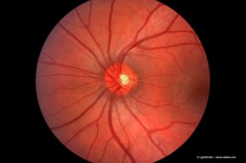
The membranes found on the retinal surface can cause visual symptoms in various conditions and a patient's vision can often be improved with surgical removal of these membranes, according to Mr Mahmut Dogramaci (The Lister Surgicentre, Lister Hospital, Stevenage, UK).
"Changing the direction at which the epiretinal membrane edge is pulled significantly affects the distribution of maximum shear stress (MSS) in the model," said Mr Mahmut Dogramaci (The Lister Surgicentre, Lister Hospital, Stevenage, UK) when he discussed the results of his studying examining the dynamics of epiretinal membrane removal off the retinal surface using a computer simulation.1
In explaining the reasoning behind the study, Mr Dogramaci stated that the membranes found on the retinal surface can cause visual symptoms in various conditions and a patient's vision can often be improved with surgical removal of these membranes.2–5 "However, very little is published about the optimum direction at which the edge of the membrane should be pulled to assist safe and effective removal," he added.
Dynamics analysis
"We identified the value and the distribution of the maximum shear stress (MSS) in the model during the membrane removal manoeuver," said Mr Dogramaci. The MSS can be used to determine the likelihood of extracellular materials failing (tearing). "We also studied its relation to the direction at which the edge of the membrane is pulled," he added.
The results of the study demonstrated that the direction at which the epiretinal membrane edge is pulled affects the distribution of the MSS and Mr Dogramaci explained that the direction was quantified by the displacement angle (DA) values in degrees. The study showed that changing the pulling direction to about 165° DA allowed the MSS to be redistributed within the tissue complex, making the removal safer and more effective. The study also showed that mean MSS value was significantly higher at the attachment pegs at all directions of pulling, proving the attachment pegs to be the most likely site of tear.
Limitations and further studies
"Computer models are widely used in engineering, less commonly used in medical and surgical, this is mainly due to the difficulties in using and understanding the basics of simulations," stated Mr Dogramaci. He explained that these limitations and difficulties could be overcome through the development of more user-friendly software that includes more biological material property data. "Further studies are now being performed to use reverse engineering techniques to find explanations for some unwanted surgical outcomes after retinal surgeries, for example, unintentional retinal shifts after retinal detachment repair," he added.
Mr Dogramaci concluded, "In reality, experienced surgeons know that changing the direction, at which the epiretinal membrane edge is pulled, significantly affects the distribution of the MSS in the surgical field and they continuously change the direction of the displacement vector as the attachment pegs give away to obtain the optimum result. Experienced surgeons learn this through self intuition. This study enabled us to explain the theory and hence facilitates teaching and learning."
References
1. M. Dogramaci and T.H. Williamson, Br. J. Ophthalmol., Published Online First 11 July 2013, doi: 10.1136/bjophthalmol-2013-303598.
2. Y.N. Hui et al., Arch. Ophthalmol., 1988;106:1280–1285.
3. R.G. Michels, Ophthalmology, 1984;91:1384–1388.
4. M.T. Trese, D.B. Chandler and R. Machemer, Graefes Arch. Clin. Exp. Ophthalmol., 1983;221:12–15.
5. S.M. Ghazi-Nouri et al., Br. J. Ophthalmol., 2006;90:559–562.
Newsletter
Get the essential updates shaping the future of pharma manufacturing and compliance—subscribe today to Pharmaceutical Technology and never miss a breakthrough.


