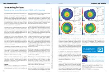
Broadening the benefits of iCare EIDON retinal imaging with the new Ultra-Wide Field Module
This article is produced by iCare Finland for healthcare professionals and is not intended to provide medical advice and/or treatment guidance. This website is not country-specific and therefore may contain information which is not applicable to your country.
By Giuseppe Querques, MD, PhD
The ability to visualize the peripheral retina is important in the diagnosis, management, and prognosis of a number of retinal diseases. By enabling early disease detection and providing information to guide initial management and follow-up decisions, identification of pathology in the retina periphery has value for optimizing clinical care.
Ultra-wide field (UWF) imaging technology offering a field of view extending to the far peripheral retina is available from several manufacturers. Recently, a UWF module became available for the iCare EIDON (Padova, Italy), a white LED confocal device. Applicable to all of the platform’s modalities – TrueColor, infrared, fundus autofluorescence, and fluorescein angiography (static imaging and dynamic videos) – the new UWF module can image up to 120° of the fundus in a single capture and up to 200° using the system’s automontage feature (SmartMosaic). The UWF module upgrade expands the benefits of imaging with the EIDON and makes it the only noncontact device in the market that can run in a fully automated mode and acquire high quality, true color confocal images of up to 200° without pupil dilation and regardless of the presence of cataract or other media opacities.
Benefits of the EIDON
By combining a confocal engine with a white light LED source, the EIDON generates images that are both rich in detail and natural in color. Analysis of the high resolution, true color images can reveal pathology that may not be evident on images acquired with non-confocal fundus cameras or on pseudocolor images generated by systems based on scanning laser ophthalmoscopy. Imaging with the EIDON thereby supports early disease detection and the opportunity for intervention when pathology may be most amenable to successful treatment.
The confocal true color EIDON images also enhance the quality of follow-up monitoring and care by improving identification of changes that signal disease progression or show response to treatment.
Images from fundus photography have a critical role in guiding treatment decisions and the confocal true color images from the EIDON platform provide a natural perception of the retina’s appearance that represents vital information in my daily practice. True color imaging provides an accurate perception of fundus anatomy while confocal technology delivers benefits of increased image sharpness, better optical resolution, and greater contrast compared to traditional fundus camera imaging.
With the benefit of true color imaging, I am confident in my ability to discriminate between normal tissue and potential signs of pathology. For example, in eyes with age-related macular degeneration, TrueColor imaging allows me to differentiate hemorrhage from pigment or to distinguish drusen from exudates.
As another important feature, the EIDON makes retinal imaging easy for the practice and the patient. The system is very simple and intuitive to use and can be operated without the need for a highly skilled photographer. In addition, image capture with the EIDON can be done regardless of the presence of media opacities and through pupils as small as 2.5 mm in diameter. The latter capability is important considering the potential for poor pupil dilation among certain patients with retinal pathology, such as those with diabetes or uveitis.
Because it is done without requiring mydriasis, imaging with the EIDON also has timesaving advantages for the practice and the patient while simultaneously improving patient comfort and satisfaction. Workflow efficiency and patient comfort are further enhanced by the speed of the imaging. With the SmartMosaic technology, images encompassing a field of view up to 200° are taken automatically in just 1 minute. With its intuitive interface, the device can be easily switched to manual mode if the user prefers.
The EIDON UWF Imaging Module
All existing EIDON devices can be upgraded with the UWF module. The UWF imaging is done with a noncontact lens that is very light (only 34 g) and fitted with a protective cap, which makes its handling safe and easy.
UWF imaging is a tremendous asset for allowing the detection of subtle signs of pathologies in the peripheral retina that are not visible with standard imaging systems. This enhanced capability for identifying abnormalities makes me more confident with my diagnosis, in my treatment decisions, and when counseling patients about management and prognosis.
For example, identification of pathologic changes in the retina periphery is important for early detection of diabetic retinopathy (DR) and for DR management. UWF imaging has been shown to be at least as sensitive and specific as the ETDRS seven standard field (SSF) montages for screening for DR. Thanks to the high resolution of its confocal true color images, UWF imaging with the EIDON has been particularly helpful to me for detecting and evaluating hemorrhages and microvascular lesions in patients with DR (PDR). The case presented in Figure 1 is from a patient with widespread proliferative DR. TrueColor Smart Mosaic allows a clear view of the complete pan-photocoagulation (Figure 1A). Looking at the central frame of the mosaic, one can see more details (Figure 1B). A tractional preretinal fibrosis along the inferior temporal arcade with neovessel and bleeding in its proximity is well visible. Pre-retinal neovascularization and epiretinal membrane are distinctly visualized. Also note the prominent perifoveal microaneurysms.
Application of the UWF module to the FA mode of the EIDON has also been valuable for evaluating and monitoring eyes with PDR. Fluorescein angiography is a routine component for follow-up of eyes with PDR, and compared to standard SSF imaging, UWF-FA allows for better detection of peripheral pathology, revealing peripheral nonperfusion, retinal neovascularization and peripheral lesions.
Figure 2A shows the FA performed on the same patient discussed above. Extensive leakage into the retina and vitreous from neovascularization elsewhere (NVE) is clearly visible in the late phase of the exam. Cystoid macular edema is also present. UWF also allows confirmation of complete ablation of ischemia in the peripheral retina.
Figure 2B presents a 9-shot montage image from a patient with non-proliferative DR. UWF imaging allows clear observation of peripheral ischemia and late peripheral venous leakage in the inferior temporal quadrant. This sign would have been missed relying upon ETDRS SSFs. In addition, the clarity of the static FA images and videos acquired with the EIDON UWF module also makes it easy to analyze results from serial examinations and determine whether there is evidence of DR progression or regression in response to therapy.
The UWF module is also useful in the evaluation of other pathologies that involve the peripheral retina, including ocular tumors, retinal tears, ischemic conditions, and degenerative disorders, such as peripheral exudative hemorrhagic chorioretinopathy in which polyps are frequently located in the infero-temporal quadrant and between the equator and the ora serrata. The case presented in Figure 3 shows the potential of the EIDON UWF module. This patient presented with a transient vitreoretinal traction following acute blunt trauma associated with incomplete posterior vitreous detachment. The patient reported a sudden onset of photopsia and floaters after the trauma. The TrueColor montage allows visualization of a tiny hemorrhage in the superior temporal quadrant at the site of the vitreoretinal traction. Although the lesion is in the extreme retinal periphery, a high-resolution image can be obtained using EIDON confocal technology.
Conclusion
UWF imaging has an important role in the evaluation and management of a number of retinal disorders, and we can expect that its applications will continue to grow as studies using this technology further our understanding of pathologies involving the peripheral retina.
With its ease of use, efficiency, and unsurpassed image quality, the EIDON has been an indispensable diagnostic tool in my practice. The UWF module augments the utility of the EIDON and my ability to provide high quality care for patients with retinal disease.
--
Giuseppe Querques, MD, PhD
Professor Giuseppe Querques is Head - Medical Retina & Imaging Unit, Department of Ophthalmology, University Vita Salute, Milan, Italy. He is an advisor to iCare.
Newsletter
Get the essential updates shaping the future of pharma manufacturing and compliance—subscribe today to Pharmaceutical Technology and never miss a breakthrough.


