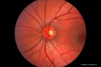
Bilateral macular changes associated with topiramate treatment
A rare case of bilateral pigmented atrophic changes in the macular region associated with topiramate treatment for chorioathetosis in a young female is reported.
Topiramate was effective in 78% of patients of resistant epilepsy.2 It is also successful in the treatment of migraine headaches, depression and bipolar disorders.3 It is a sulpha-derivative and acts by blocking glutamate receptors, enhancing the effect of gamma amino butyric acid neurotransmitters and inhibition of Kainate mediated conductance at glutamate receptors of the AMPA/Kainite type.4 Its chemical structure is 2,3:4,5-bis-O-(menthylethylidene)-B-D fructopyranose sulphamate.5
A fourteen-year-old Caucasian female was referred by her optician to the eye outpatient clinic. He noted, during a routine regular yearly check up, pigmentary changes at both maculae. The same optician had not noted these changes on previous visits.
She had been on topiramate for the treatment of Familial Paroxysmal Chorioathetosis 75 mg (am) and 100 mg (pm) a day for a year prior to attending her optician when the macular changes were noted.
On examination the best-corrected visual acuity was 6/6 in both eyes, amsler chart test was normal and colour vision (Ischihara) was perfect. Humphrey visual fields did not show any abnormality and the anterior segment examination was unremarkable, as it demonstrated normal intraocular pressures. Fundal examination revealed symmetrical atrophic and pigmentary changes in the macular regions between the fovea and the optic disc. The rest of the retinal examination was not unusual.
Fluorescein angiogram showed window defects corresponding to the atrophic changes with no leakages. The rest of the choroid, retinal vasculature, The ERG and EOG were normal.
Newsletter
Get the essential updates shaping the future of pharma manufacturing and compliance—subscribe today to Pharmaceutical Technology and never miss a breakthrough.


