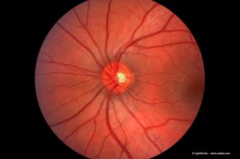
Advanced quantitative imaging can potentially improve screening for hydrochloroquine retinopathy
Non-invasive techniques may detect signs of drug-induced toxicity at a pre-clinical stage
In a poster presented at the Retina Congress, Dr Kimberly Stepien, assistant professor of ophthalmology at the Institute and her colleagues reported on findings from imaging performed with spectral-domain optical coherence tomography (SD-OCT) and adaptive optics (AO) in two patients with evidence of hydroxychloroquine retinopathy based on clinical examination.
Detecting toxicity
"Discontinuing hydroxychloroquine at the earliest sign of retinal toxicity is important for preserving vision," she said. "Although retinopathy can continue to progress after hydroxychloroquine is stopped, there are multifocal ERG data suggesting that early toxicity can be reversible with prompt cessation of the drug.
"Therefore, the sooner hydroxychloroquine toxicity is detected, the sooner treatment can be stopped to try to preserve vision," she said. "Our experience with SD-OCT and AO imaging is limited, and further research is needed.
"However, these techniques are showing exciting potential for identifying hydroxychloroquine retinopathy at a 'pre-clinical' state and simultaneously offer a number of additional advantages compared with current screening methods that make them attractive monitoring tools," she added.
Currently, the American Academy of Ophthalmology Preferred Practice Patterns for screening for hydroxychloroquine retinopathy recommend an annual comprehensive eye exam and testing with either perimetry (10-2 Humphrey Visual Field Analyzer, Carl Zeiss Meditec) or an Amsler grid. Use of additional imaging is left to the discretion of the provider.
"Extensive retinal damage has to occur before retinopathy can be detected by these testing methods, and fundoscopic changes tend to be a late finding," Dr Stepien said. "Furthermore, Amsler grid testing can be very subjective, and visual field testing has a learning curve, with many tests being inconclusive.
"Other modalities, including multifocal ERGs, have shown promise," she said. "However, this test is expensive, requires special instrumentation as well as special training for administration and interpretation and is not usually available in the clinic, so that a second appointment is required."
Newsletter
Get the essential updates shaping the future of pharma manufacturing and compliance—subscribe today to Pharmaceutical Technology and never miss a breakthrough.


