
|Articles|November 1, 2006
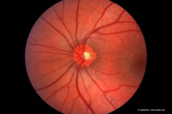
Retinal vein occlusion therapy: can we reach a consensus?
Retinal venous occlusions (RVO) are the most common visually disabling retinopathy after diabetes and age-related macular disease.1 The pathogenesis of RVO is multifactorial with both local factors and systemic diseases being etiologically important. It is essential that each individual contributing mechanism is established so that a beneficial treatment protocol can be developed to treat this debilitating condition.
Advertisement
A definite pathogenic event in all cases is the development of intralumenal thrombus, which may arise from a combination of four primary mechanisms: degenerative vessel wall changes, abnormal coagulation factors, abnormal blood flow, and apposition of sclerotic vessels in a common adventitial sheath.2
A number of reports have described the localizing influence of anterior-artery-to-vein crossings in eyes with branch RVO and an anatomical study confirmed focal venous constriction at these sites in non-hypertensive individuals. The ocular axial length in central retinal vein occlusion (CRVO) and branch retinal vein occlusion (BRVO) were significantly shorter than in controls.3 This significant difference may be an additional risk factor for CRVO and BRVO. Many studies including case control studies have also examined the clinical features and risk factors in younger and older subjects.
There are still gaps in understanding the aetiology and pathogenesis of circulatory disorders of the retinal veins. Although various new therapeutic approaches have been developed in the past few years, existing therapy forms are subject to controversy and available data is, to some extent, inconsistent. The objective of this review is to evaluate the treatments commonly advocated, in the light of our current scientific knowledge of RVO.
Intravitreal drug administration
Intravitreal thrombolysis
This therapy for RVO aims to dissolve the preformed thrombus. Several, largely retrospective, studies have been performed on the benefits of intravitreal thrombolysis in this setting, but opinions still vary. While some researchers report a variable amount of improvement in visual acuity (VA), others report no beneficial effect, while others still consider this treatment approach to be too risky.
Specifically, treatment with the fibrinolytic agent intravitreal tissue plasminogen activator (tPA) has been assessed with varying outcomes. While teams led by Elman6 and Ghazi7 reported improvements in VA with the agent, Weizer and Fekrat8 suggested tPA may be a useful treatment for CRVO with a duration of 21 days or less. In contrast, Glacet-Bernard found that it did not significantly modify the course of the disease and the final visual outcome did not differ significantly from that observed in the natural course of the disease.9
In a case report, Lam and Blumenkranz12 performed vitrectomy with lysis of vitreopapillary and epipapillary adhesions, subretinal peripapillary tPA injection and photocoagulation, and claimed that this combined procedure 'appeared to accelerate the resolution' of CRVO.
Newsletter
Get the essential updates shaping the future of pharma manufacturing and compliance—subscribe today to Pharmaceutical Technology and never miss a breakthrough.
Advertisement
Related Content
Advertisement


Latest CME


Advertisement
Advertisement