
News|Videos|November 21, 2023
AAO 2023: Small aperture IOL implantation and retinal visualisation
Mark Blecher, MD, spoke with the Ophthalmology Times team about his poster addressing retinal visualisation and patients who have undergone small aperture IOL implantation at this year's American Academy of Ophthalmology meeting
Advertisement
Mark Blecher, MD, spoke with the Ophthalmology Times team about his poster addressing retinal visualisation and patients who've had small aperture IOL implantation at this year's American Academy of Ophthalmology meeting.
Video Transcript
Editor's note - This transcript has been edited for clarity.
Mark Blecher, MD:
Good morning from the American Academy of Ophthalmology meeting in San Francisco. I'm Mark Blecher. I'm an attending surgeon at Wills Eye Hospital, and I have the honor and pleasure to be presenting a poster here at the meeting, addressing retinal visualisation and patients who've had small aperture IOL implantation.
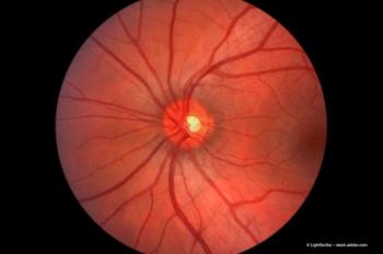
This paper derives from a small subset of the IC-8 Apthera, a small-aperture IOL FDA clinical trial study that was conducted a couple of years ago. The lens has now been approved by the FDA. And this small subset of patients in that study were utilised to look at any secondary, knock-on issues related to having a small aperture IOL in the eye.
Specifically, in this particular case, peripheral retinal visualisation, posterior fundus visualisation and analysis with OCT, and visual field testing. In this small group of the original of 49 eyes, we were taking fundus photographs. They were looked at and evaluated by both the surgeons and by an independent mass-blinded reading center. The results were that of all the peripheral fundus photos and the OCTs, there was not noted to be any difference in utilisation of the test or quality of the exam itself between control eyes with a standard monofocal lens and the IC-8 Apthera small aperture IOL.
Visual field studies were a little more difficult to assess, but if you know anything about visual fields, you need a much larger in to evaluate deviations in visual field from one test to another. So it did not seem that there was a notable difference between the groups, but I'd hesitate to imply any clinical–any clinical significance to that without a larger study. In summary, we don't feel that the use of the IC-8 small aperture IOL should impede the visualisation of the retina, either peripherally or centrally, with either fundus photography or OCT.
Newsletter
Get the essential updates shaping the future of pharma manufacturing and compliance—subscribe today to Pharmaceutical Technology and never miss a breakthrough.
Advertisement
Related Content
Advertisement


Latest CME


Advertisement
Advertisement