
|Articles|June 1, 2007
AMD: combining therapies = combined synergies?
Pilot study, which examines the benefits of combining photodynamic therapy (PDT) with bevacizumab (Avastin) in AMD patients, yields promising results.
Advertisement
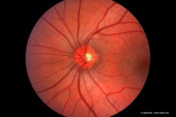
Two currently available treatments include photodynamic therapy (PDT) and anti-angiogenic therapy. In terms of its mechanism of action, PDT involves the photosensitive dye, verteporfin, which is administered intravenously. It circulates and accumulates in the abnormal blood vessels caused by neovascularization and is activated with non-thermal laser application of light generating free radicals. These reactive oxygen products then selectively close the abnormal blood vessels by damaging the endothelium.
Anti-angiogenic drugs, on the other hand, are the newest class of agents to be approved and act by inhibiting new vessel growth through vascular endothelial growth factor (VEGF) inhibition; the stimulus for new vessel development and fluid leakage. The actions of both agents are temporary.
Administering a good combination?
In order to put the theory to the test, we performed a pilot study, treating 12 patients with CNV resulting from AMD with a treatment regimen of PDT followed by an intravitreal injection of the off-label anti-VEGF agent bevacizumab.
In terms of the treatment protocol, PDT was performed (following the approved protocol) and this was followed five to seven days later by a 2.5 mg intravitreal injection of bevacizumab, which was administered under sterile conditions.
Patients were followed-up at one, three and six months post-therapy, when a fluorescein angiogram (FA) and optical coherence tomography (OCT) was performed. If visual acuity (VA) had decreased by more than one line and/or OCT showed a greater than 10% increase in retinal thickness from baseline, another intravitreal injection was administered at follow-up. No patient received additional PDT and statistical analysis was undertaken using Wilcoxon signed rank test.
All patients had a best corrected visual acuity (BCVA) of between 20/40 and 20/400. A full ophthalmic examination was performed, as well as a baseline FA and OCT. The baseline average VA was 20/80 (logMAR 0.63±0.18) and average foveal thickness was 343±63 μm.
How do these results compare with others?
Foveal thickness also improved to 247 μm at one month and continued to decrease up to three and six months (231±57.7 μm). This compares favourably with the recently presented P.R.O.J.E.C.T. study results.
Newsletter
Get the essential updates shaping the future of pharma manufacturing and compliance—subscribe today to Pharmaceutical Technology and never miss a breakthrough.
Advertisement
Related Content
Advertisement


Latest CME


Advertisement
Advertisement